Premium USA-Made Research Compounds
Browse lab-tested peptides, research liquids, capsules and more.
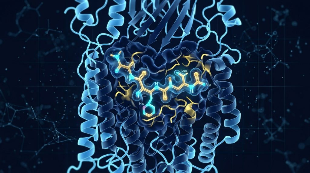
IGF-1 DES (Truncated IGF-1): Receptor Binding Kinetics, Potency Research & Laboratory Applications
Among the growing catalog of insulin-like growth factor analogs studied in preclinical research, IGF-1 DES occupies a distinctive position. This truncated variant — formally designated des(1-3)IGF-1 — differs from native IGF-1 by just three amino acids at its N-terminus, yet that subtle structural difference produces a markedly altered biochemical profile. For researchers investigating the GH-IGF axis, anabolic signaling cascades, or tissue-specific growth factor dynamics, understanding why this truncation matters at the molecular level is essential.
What Is IGF-1 DES? The Structural Basis of a Truncated Analog
Native insulin-like growth factor-1 (IGF-1) is a 70-amino-acid polypeptide that shares structural homology with proinsulin. IGF-1 DES is the result of removing the first three residues — glycine, proline, and glutamate (Gly-Pro-Glu) — from the N-terminal end of the full-length molecule. This truncation arises naturally in certain tissues, most notably in the brain and gastrointestinal tract, where local proteolytic processing generates the des(1-3) form endogenously.
Why does this matter to researchers? The loss of just three residues fundamentally reorganizes the peptide’s interaction landscape. The N-terminal tripeptide in native IGF-1 is not merely structural filler — it forms part of a critical binding interface with insulin-like growth factor binding proteins (IGFBPs). Remove it, and the peptide’s relationship with its carrier proteins changes dramatically.
Looking for Premium Research Compounds?
Structurally, the remainder of the molecule is preserved. The three-disulfide bond framework, the receptor-binding domains, and the C-domain architecture remain intact. IGF-1 DES can still engage the IGF-1 receptor (IGF-1R) and the insulin receptor (IR) with high affinity. What changes is not the peptide’s capacity to signal — it is the peptide’s freedom to do so.
The IGFBP Binding Story: Why Truncation Amplifies Bioavailability
This is the central research insight behind IGF-1 DES. In circulating blood and interstitial fluid, the vast majority of native IGF-1 is bound to one of six insulin-like growth factor binding proteins (IGFBP-1 through IGFBP-6). These carrier proteins serve multiple functions: they extend the half-life of IGF-1, modulate its distribution, and critically, prevent premature receptor engagement. Only a small fraction of native IGF-1 exists in a “free” form capable of activating IGF-1R.
The truncation in IGF-1 DES dramatically reduces affinity for IGFBPs — by approximately 70-fold compared to full-length IGF-1. This is not a marginal reduction. It is a near-complete decoupling of the peptide from its most prominent regulatory constraints.
What are the downstream consequences for researchers? Because IGF-1 DES does not sequester into IGFBP complexes at the same rate, a far greater proportion of the peptide remains “free” and available for receptor engagement at any given moment. In vitro studies have consistently demonstrated that IGF-1 DES elicits receptor activation responses at concentrations far below those required to achieve equivalent effects with native IGF-1. Some comparative binding studies report potency differentials of 10- to 100-fold depending on the cell type and assay conditions used.
This dynamic has important implications for experimental design. When researchers model IGF-1R activation in cell culture — where no systemic IGFBP reservoir exists — native IGF-1 and IGF-1 DES behave more similarly. But in ex vivo or in vivo systems where IGFBPs are present, the DES analog’s reduced sequestration becomes a decisive variable.
Receptor Binding Kinetics: Potency at the IGF-1R Interface
The IGF-1 receptor (IGF-1R) is a receptor tyrosine kinase whose activation initiates the PI3K/Akt and MAPK/ERK signaling cascades — pathways involved in cell survival, proliferation, differentiation, and metabolic regulation. Understanding how IGF-1 DES interacts with this receptor, compared to native IGF-1 and its synthetic analog IGF-1 LR3, is a key area of active inquiry.
Receptor binding studies have shown that IGF-1 DES retains strong affinity for IGF-1R. Some reports suggest its intrinsic receptor-binding affinity is marginally lower than native IGF-1 when measured in isolation — but this is more than compensated by the dramatically elevated free-peptide fraction that results from reduced IGFBP binding in biological systems. The net effect is higher effective potency.
Kinetic studies examining association and dissociation rates at IGF-1R have noted that the truncated N-terminus does not appear to substantially alter the receptor’s ligand-binding domain recognition. The primary binding interface, involving the B- and C-domains of IGF-1, remains structurally intact in des(1-3)IGF-1. This suggests that receptor activation dynamics — once binding occurs — closely mirror those of the full-length peptide.
Downstream signaling studies in myoblast and neuronal cell models have corroborated this. Phosphorylation of Akt (Ser473) and ERK1/2 following IGF-1 DES stimulation follows kinetics consistent with robust IGF-1R engagement, with observed EC50 values in cell culture contexts reflecting the peptide’s enhanced free-fraction availability.
IGF-1 DES vs. IGF-1 LR3: Two Approaches to the Same Problem
Researchers frequently compare IGF-1 DES to its synthetic cousin, IGF-1 LR3. Both analogs were designed — in different ways — to extend or amplify IGF-1 bioactivity by reducing IGFBP-mediated sequestration. The mechanisms, however, are fundamentally different.
IGF-1 LR3 is a full-length 83-amino-acid analog that retains the intact N-terminus but incorporates an arginine substitution at position 3 (replacing glutamate) and an N-terminal 13-amino-acid extension. These modifications significantly reduce IGFBP-3 and IGFBP-5 binding affinity, resulting in an extended half-life in biological systems — typically estimated at several hours in vivo, compared to the much shorter half-life of native IGF-1 or IGF-1 DES.
IGF-1 DES takes the opposite structural approach. Rather than extending the sequence, it shortens it. The consequence is a peptide with a shorter half-life than LR3, but a more acute and localized receptor activation profile. Where LR3 is designed for sustained systemic exposure, DES is characterized by fast, high-potency, localized action.
Which analog is more appropriate for a given research question? It depends entirely on the experimental model. Studies examining acute signaling events or tissue-localized receptor dynamics may favor the DES analog. Research requiring prolonged receptor engagement or systemic distribution modeling may favor LR3. Many researchers have employed both in parallel to deconvolute the temporal components of IGF-1 signaling.
Key Research Applications: Where IGF-1 DES Has Been Studied
The biochemical profile of IGF-1 DES has made it a useful tool across several research domains.
Muscle Satellite Cell Activation and Myoblast Differentiation
Skeletal muscle research has long used IGF-1 analogs to investigate satellite cell biology. Satellite cells — the resident stem cells of skeletal muscle — require IGF-1 signaling for activation and differentiation following stress or injury. IGF-1 DES has been studied in myoblast cultures to examine its capacity to stimulate proliferation and differentiation, with findings supporting potent activation of the IGF-1R-PI3K-Akt axis at low nanomolar concentrations. These studies provide mechanistic insight into anabolic signaling in muscle tissue at the molecular level.
Neural Tissue Growth Factor Research
The endogenous presence of des(1-3)IGF-1 in brain tissue has made it a natural focus of neurotrophic research. IGF-1 DES has been studied in neuronal cell models for its neuroprotective signaling potential, with investigations focusing on ERK-mediated survival signaling and differentiation-promoting effects in various neuronal lineages. Its endogenous occurrence in the CNS lends these studies physiological relevance.
Bone Matrix and Osteoblast Studies
Bone biology researchers have investigated IGF-1 DES in osteoblast models, examining its effects on extracellular matrix synthesis and mineralization marker expression. Given that IGF-1 signaling is integral to bone formation and remodeling, the truncated analog serves as both a mechanistic probe and a comparative tool against full-length IGF-1.
GH-IGF Axis Research
Understanding how local versus systemic IGF-1 variants modulate GH-IGF axis feedback is an important area of endocrine research. IGF-1 DES — as a naturally occurring, locally produced form of IGF-1 — provides researchers with a tool to investigate autocrine and paracrine IGF-1 signaling independently of systemic IGFBP-regulated circulating IGF-1.
Conclusion
IGF-1 DES represents a structurally elegant research tool whose value derives directly from its simplicity: remove three amino acids, and the peptide’s regulatory constraints are fundamentally altered. The ~70-fold reduction in IGFBP binding affinity transforms the peptide from a heavily sequestered, tightly regulated signaling molecule into a free, rapidly acting receptor agonist with concentrated local potency.
For researchers designing studies around IGF-1 receptor kinetics, anabolic signaling cascades, tissue-specific growth factor responses, or comparisons within the broader IGF axis, the DES analog offers a uniquely informative biochemical profile. Its contrast with IGF-1 LR3 — both addressing IGFBP sequestration but through opposite structural strategies — makes it particularly valuable in studies requiring temporal or mechanistic differentiation of IGF-1 signaling effects.
As research into IGF-1 system modulation continues to advance, des(1-3)IGF-1 remains a foundational reference compound for understanding the interplay between structural conformation, carrier protein dynamics, and receptor-level bioactivity in the insulin-like growth factor system.
For Research Purposes Only: The information presented in this article is intended solely for scientific research and educational purposes. These compounds are not approved for human use and should only be handled by qualified researchers in appropriate laboratory settings in compliance with all applicable regulations.
Continue Your Research
Explore our complete catalog of premium research compounds.