Twenty-eight amino acids. That’s all it takes. Vasoactive Intestinal Peptide : VIP, as it’s universally abbreviated in the literature : is a neuropeptide of almost disarming simplicity in structure, yet its functional reach spans the immune system, the central and peripheral nervous systems, the gut, the lungs, and the body’s internal clock. Researchers who first characterized VIP in the early 1970s from porcine intestinal tissue could scarcely have anticipated just how far that thread would unravel. Decades on, VIP remains one of the most intellectually compelling peptides in biomedical research, precisely because it refuses to stay in one lane.
Structure and Distribution: A Small Peptide with a Large Presence
VIP belongs to the glucagon/secretin superfamily of peptides : a structural grouping that includes PACAP, secretin, glucagon, and GIP, among others. Its 28-amino-acid sequence is highly conserved across mammalian species, which itself signals functional importance. Evolution doesn’t tend to preserve what doesn’t matter.
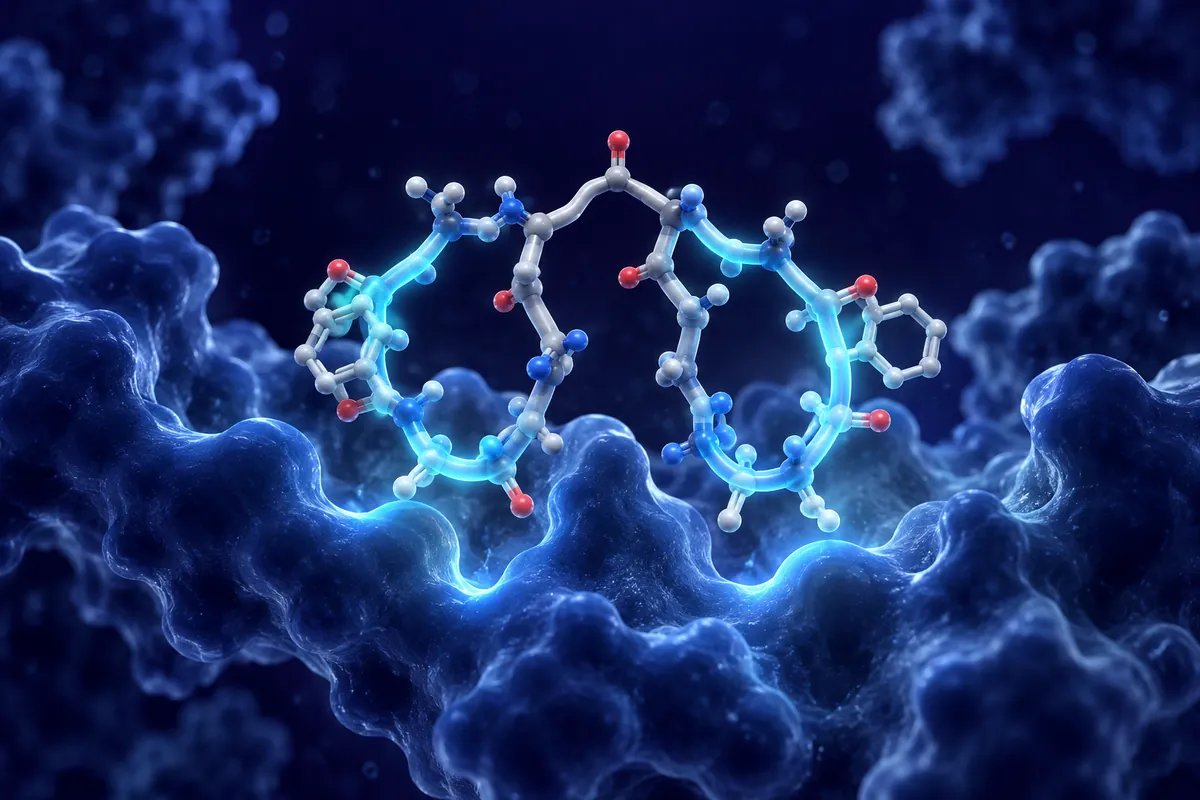
The peptide is synthesized from a larger precursor protein, prepro-VIP, which also encodes peptide histidine methionine (PHM-27) and peptide histidine isoleucine (PHI-27), depending on the species. VIP’s α-helical conformation in solution : particularly its C-terminal amphipathic helix : is central to how it interacts with its cognate receptors. Structure, here, is not incidental to function; it is function.
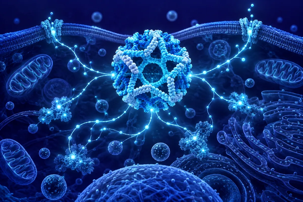
Where is VIP found? Almost everywhere that matters. It’s expressed abundantly in the enteric nervous system, in hypothalamic neurons (particularly the suprachiasmatic nucleus), in pulmonary tissue, in immune cells including T cells and mast cells, and in parasympathetic nerve terminals throughout the cardiovascular system. This near-ubiquitous distribution is less a redundancy and more a statement: VIP is woven into the architecture of multiple regulatory systems simultaneously.
Receptor Pharmacology: VPAC1, VPAC2, and the PAC1 Question
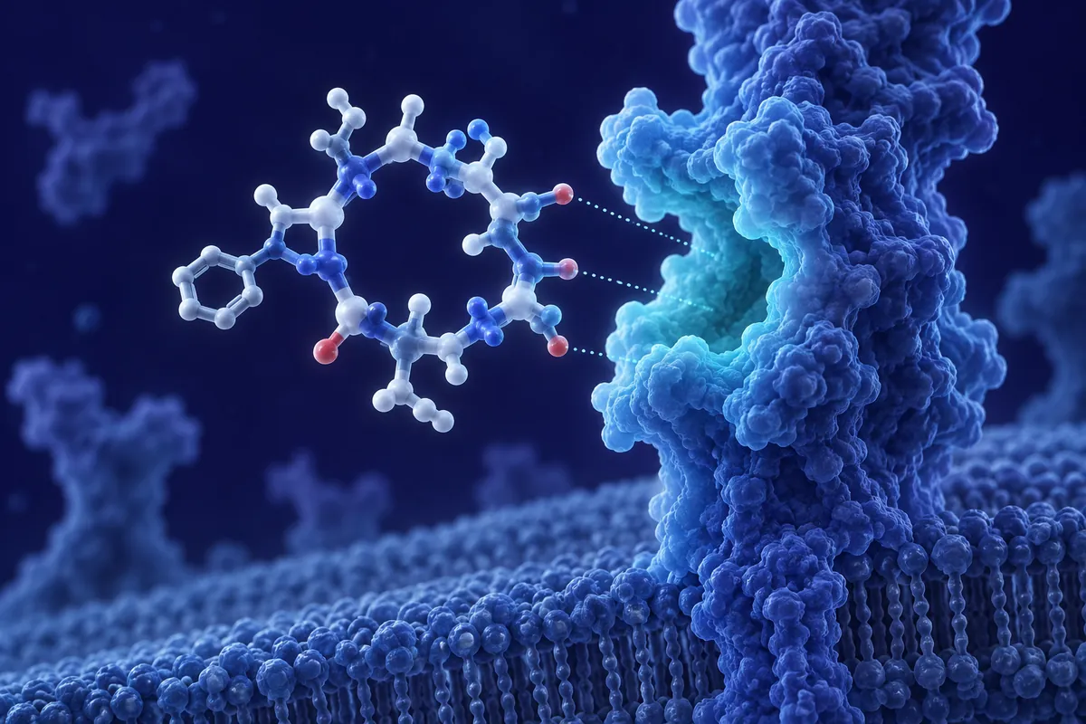
VIP exerts its effects through two primary G protein-coupled receptors : VPAC1 (encoded by VIPR1) and VPAC2 (encoded by VIPR2), both of which couple primarily to Gαs and drive adenylyl cyclase activation, elevating intracellular cAMP. VIP also binds PAC1, the primary receptor for PACAP, though with substantially lower affinity. This receptor promiscuity is worth noting : it means VIP’s signaling landscape can shift depending on which receptor subtypes are expressed in a given tissue.
VPAC1 is expressed broadly, particularly in lung, liver, and immune cells. VPAC2 shows a more selective distribution : prominently in the suprachiasmatic nucleus (SCN), smooth muscle, and certain immune subpopulations. This receptor-level segregation is one reason VIP can do such different things in different tissues without producing contradictory outcomes. Researchers studying context-specific effects will often examine which receptor subtype predominates in their model system before drawing broader conclusions.
Downstream Signaling Complexity
cAMP elevation is the canonical VIP signal, but the picture downstream is considerably richer. Depending on cell type, VPAC activation can engage PKA, EPAC (exchange protein directly activated by cAMP), MAPK pathways, and PI3K/Akt signaling.
If there’s one area of VIP research that has attracted particularly intense investigation over the past two decades, it’s immunomodulation. But the mechanistic details are where the story gets interesting.
Research by Delgado, Ganea, and colleagues has been particularly instrumental in mapping these circuits : their work through the early 2000s established much of what we now take as foundational in VIP immunobiology.
One especially active area involves regulatory T cells (Tregs).
Mast Cells, Neuropeptides, and the Neuroimmune Interface
VIP is also notable at the neuroimmune interface : that conceptually rich border where the nervous system and immune system communicate. Mast cells express VIP receptors and release their own VIP upon activation, creating a potential autocrine/paracrine loop. Parasympathetic nerve fibers in lymphoid tissues release VIP in proximity to immune cells. The question of how, precisely, neural VIP release translates into immune outcomes in vivo remains an active research puzzle : one that is difficult to study cleanly but carries significant implications for understanding stress-immune interactions.
Circadian Biology: VIP as the Clock’s Timekeeper
Here is perhaps VIP’s most fascinating role. The suprachiasmatic nucleus : the brain’s master circadian pacemaker : contains a dense population of VIP-expressing neurons, and VIP/VPAC2 signaling is now understood to be essential for synchronizing cellular oscillators within the SCN network itself. This is not a peripheral or modulatory role. It’s central.
Individual SCN neurons each harbor molecular clock machinery : the CLOCK/BMAL1/PER/CRY feedback loops that generate ~24-hour rhythms at the cellular level. But without intercellular synchronization, these individual oscillators drift apart. VIP, released from a subset of SCN neurons in a rhythmic, light-responsive manner, coordinates the phase and amplitude of the network. Mice with targeted disruptions of VIP or VPAC2 exhibit profound disruptions in circadian behavior : fragmented locomotor rhythms, attenuated or lost periodicity under free-running conditions.
Why does any of this matter to researchers? Because the SCN clock doesn’t just govern sleep-wake cycles. It coordinates oscillators in peripheral tissues : liver, lung, adrenal gland, immune cells : via hormonal and neural outputs. VIP’s role at the top of that hierarchy means it influences the temporal organization of physiology far beyond the hypothalamus.
VIP’s presence in the nervous system extends well beyond the SCN. It’s found in cortical interneurons, in the hippocampus, in the spinal cord, and throughout the peripheral autonomic nervous system.
Some of this appears to involve upregulation of survival-associated signaling (including BDNF and Bcl-2 family members), while some may relate to VIP’s ability to modulate glial activation states.
VIP-expressing interneurons in the cortex occupy a specific functional niche, targeting primarily inhibitory interneurons and thereby disinhibiting local circuits.
Research Outlook: One Peptide, Many Questions
What makes VIP such a compelling research target is precisely the integration problem it poses. Do VIP’s immunomodulatory effects follow a circadian pattern? Does chronic neuroinflammation affect VIP expression in the SCN and, consequently, circadian function?
These are not idle questions. Research groups across neuroimmunology, chronobiology, and neuropeptide pharmacology are beginning to ask them in earnest. The tools available : conditional knockout models, optogenetic approaches, high-resolution receptor imaging, and advanced peptide analogs with subtype-selective receptor profiles : are increasingly capable of probing the kind of system-level interactions that VIP’s biology seems to demand.
For researchers working in any of these domains, VIP offers something increasingly rare in molecular biology: a genuine integrative node.
Disclaimer: This content is intended for research purposes only and is not meant to constitute medical advice.
This educational material discusses laboratory research concepts only. Loti Labs products are intended strictly for research and development use and are not for human consumption.