Premium USA-Made Research Compounds
Browse lab-tested peptides, research liquids, capsules and more.
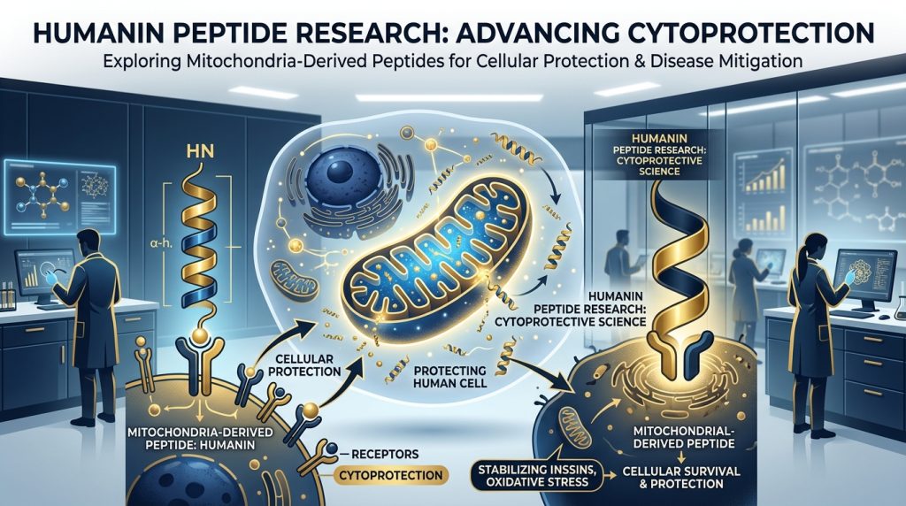
Humanin Peptide: Mitochondrial-Derived Longevity Research, IGF-1 Signaling & Neuroprotection Studies
Mitochondria have long been described as the cell’s power plants — but that framing undersells them. Over the past two decades, researchers have recognized that mitochondria are active signaling organelles, capable of producing small peptides that communicate with the rest of the cell and even with distant tissues. Humanin is one of the most studied of these mitochondria-derived peptides (MDPs), and the research surrounding it has grown into something genuinely remarkable.
Identified first in the context of neurodegeneration, humanin has since drawn attention from longevity researchers, metabolic scientists, and cardiologists alike. Its interactions with the IGF-1 axis, its neuroprotective properties in laboratory models, and its associations with healthy aging in long-lived populations all make it a molecule worth understanding in depth. What follows is a research-focused overview of what science currently knows — and where investigational work is heading. All content is intended strictly for research use only.
What Is Humanin?
Humanin is a small peptide — 21 amino acids in length — that sits at a fascinating intersection of mitochondrial biology and cellular survival signaling. It is not synthesized through the conventional nuclear genome pathway. Instead, it is encoded within the mitochondrial genome itself, specifically within the 16S ribosomal RNA gene of mitochondrial DNA. This alone makes it unusual. Most bioactive peptides trace back to nuclear-encoded genes; humanin is one of a growing class of exceptions.
Discovery and Molecular Identity
The discovery of humanin came in 2001, from the laboratory of Ikuo Nishimoto and colleagues. The team was screening for genes that could protect neurons against Alzheimer’s disease–related cell death — a challenging problem, since the precise mechanisms of neuronal loss in Alzheimer’s were (and remain) incompletely understood. When they identified a transcript from the occipital cortex of an Alzheimer’s-affected brain that conferred protection against cell death in culture models, they were surprised to trace it back to a mitochondrial locus rather than a nuclear gene. The peptide was named humanin to reflect its apparent role in preserving human neuronal survival.
Structurally, humanin’s 21-amino acid sequence (MAPRGFSCLLLLTSEIDLPVK) is highly conserved across mammals. Circulating forms of humanin have been detected in human blood and cerebrospinal fluid. Levels decline with age — a pattern that has drawn considerable interest from longevity researchers.
Humanin Analogs: HNG and Beyond
Shortly after the original identification, researchers began modifying the humanin sequence to understand its structure-activity relationships and to develop more potent analogs for laboratory investigation. The most extensively studied is HNG (humanin with a glycine-to-serine substitution at position 14). This single amino acid change — replacing glycine with serine — dramatically increases the peptide’s bioactivity, reportedly by orders of magnitude compared to the native sequence in certain assay conditions.
HNG has become a workhorse compound in humanin research precisely because its enhanced potency allows investigators to study receptor binding, downstream signaling, and functional outcomes at lower concentrations. Other analogs have also been synthesized and characterized, each offering slightly different receptor interaction profiles. For research programs focused on mechanistic studies, the analog landscape is an important context to keep in mind.
Mitochondrial Origin and the MOTS-c Connection
Humanin was not a lone discovery for long. As researchers began to appreciate that mitochondria could encode bioactive peptides, additional MDPs were identified. MOTS-c (mitochondrial open reading frame of the 12S rRNA type-c) is perhaps the closest research sibling to humanin — another small peptide with significant metabolic and longevity-related biology, also encoded in the mitochondrial genome.
The existence of MOTS-c alongside humanin reinforced a broader hypothesis: that mitochondria serve as endocrine-like organelles, releasing peptide signals in response to cellular stress, energy status, and aging. This framework has meaningfully reshaped how some researchers think about the molecular biology of aging.
Pinchas Cohen at the University of Southern California has been central to advancing this view. His laboratory’s work on humanin and MOTS-c helped establish that these peptides circulate systemically and appear to coordinate responses across tissues — they aren’t simply local mitochondrial signals. Understanding humanin as part of a broader mitochondrial signaling network, rather than in isolation, provides richer context for the research data.
One striking finding from population studies: centenarians and their offspring tend to show higher circulating humanin levels than age-matched controls. The correlation doesn’t establish causality, but it has spurred significant interest in what role humanin levels may play in exceptional longevity.
IGF-1 Signaling Pathways in Humanin Research
IGF-1, or insulin-like growth factor 1, is a central axis in aging biology. The IGF-1/insulin signaling pathway is one of the most evolutionarily conserved regulators of lifespan across species from nematodes to mammals. Humanin connects to this axis in several meaningful ways, which has made it especially interesting to researchers working at the crossroads of longevity and metabolism.
IGFBP-3 and Humanin Interactions
One of the better-characterized molecular interactions involving humanin is its binding to insulin-like growth factor binding protein 3 (IGFBP-3). IGFBP-3 is the most abundant IGF binding protein in human circulation, regulating bioavailability of IGF-1 and IGF-2 while exerting its own independent effects on cell survival and apoptosis.
Research has demonstrated that humanin binds directly to IGFBP-3, influencing its pro-apoptotic activity. In cell-based models, this interaction has been associated with attenuation of IGFBP-3-induced apoptosis — though the precise mechanisms are still being parsed. The interaction is not merely competitive; it appears to alter IGFBP-3 signaling in ways relevant to cell survival under stress.
Humanin also interacts with IGFBP-5, though this has received somewhat less mechanistic attention. Both binding proteins are implicated in growth regulation and aging, making these interactions relevant to longevity-focused research programs.
Downstream Cellular Signaling
On the receptor side, humanin has been shown to engage the formyl peptide receptor-like 1 (FPRL1, also known as FPR2), a G protein-coupled receptor with broad relevance to inflammation resolution and cell survival. Through this receptor, humanin activates downstream pathways including STAT3, PI3K/Akt, and MAPK signaling cascades — all of which play roles in cell survival, metabolism, and stress response.
The JAK2/STAT3 pathway activation has been of particular interest in neuroprotection research, since STAT3 signaling is associated with neuronal survival in several disease models. Meanwhile, PI3K/Akt activation ties humanin’s receptor biology directly to insulin signaling, providing a mechanistic link between humanin and metabolic function that goes beyond mere correlation.
Researchers have also described a trimeric receptor complex involving cytokine-like receptor 1 (CNTFR), WSX-1, and gp130 as a high-affinity humanin receptor. This complex, involved in cytokine signaling, suggests that humanin’s receptor biology is more complex than a single receptor interaction — a feature common to peptides with pleiotropic activity profiles.
Neuroprotective Mechanisms in Laboratory Models
Neuroprotection was the lens through which humanin was first understood, and it remains one of the richest areas of ongoing preclinical investigation. Laboratory models have now documented humanin’s apparent protective effects across multiple insults relevant to neurodegeneration — not just the amyloid-beta pathway that first put it on the map.
Alzheimer’s Disease Research Models
The original Nishimoto paper demonstrated that humanin could protect neurons from cell death induced by familial Alzheimer’s disease–associated genes (specifically presenilin-2 mutants and V642I APP). Subsequent work expanded that initial observation considerably. In cell culture models, humanin has shown protective activity against a range of Alzheimer’s-relevant insults, including oxidative stress, ER stress, and direct amyloid-beta toxicity.
In rodent models, intracerebroventricular administration of humanin or HNG has been associated with improvements in spatial memory and reductions in markers of neuroinflammation and oxidative damage. These are controlled laboratory observations — they illuminate mechanism and biological plausibility rather than clinical application. The consistency of the neuroprotective signal across multiple labs and model systems has nonetheless made humanin a credible focus for continued neurodegeneration research.
Nir Barzilai’s group at Albert Einstein College of Medicine has contributed substantially here, including population-level data linking humanin levels to cognitive function in aging cohorts. Such epidemiological associations complement the mechanistic work and help frame testable hypotheses for future investigational studies.
Amyloid-Beta Interaction Studies
One particularly interesting line of research concerns direct physical interaction between humanin and amyloid-beta (Aβ) peptides — the principal protein constituent of the plaques seen in Alzheimer’s-affected brain tissue. Several studies have reported that humanin can bind directly to Aβ peptides, potentially inhibiting their aggregation into the oligomeric and fibrillar forms thought to be neurotoxic.
This direct interaction hypothesis is mechanistically appealing: a small protective peptide that physically intercepts the toxic species before they can form aggregates. Whether this mechanism is relevant at physiologically relevant concentrations — and whether it operates in vivo as it does in in vitro assays — remains an important open question that future research programs will need to address.
Beyond amyloid, humanin has also shown protective activity in models of ischemia-reperfusion injury, oxidative stress–induced neuronal death, and even retinal degeneration — highlighting that its neuroprotective biology is not narrowly confined to Alzheimer’s-related mechanisms.
Longevity and Anti-Aging Research
The connection between humanin and longevity is one of the more compelling threads in the entire humanin research literature. It is not simply that the peptide is anti-apoptotic or cytoprotective — those properties alone wouldn’t distinguish it from dozens of other survival factors. What makes the longevity connection interesting is the convergence of population data, model organism findings, and mechanistic overlap with established longevity pathways.
C. elegans and Mammalian Aging Models
C. elegans — the tiny roundworm that has been central to aging biology since the 1990s — has also served as a platform for studying humanin. Work in C. elegans models has shown that humanin administration can extend lifespan, and that this effect appears to involve the insulin/IGF-1 signaling pathway, particularly through DAF-16, the worm ortholog of the FOXO transcription factor family.
FOXO transcription factors are central regulators of longevity across many species. The fact that humanin’s lifespan-extending effects in worms appear dependent on this pathway places it squarely within established longevity biology rather than as an outlier phenomenon. This kind of evolutionary conservation is taken seriously by aging researchers — mechanisms that matter in worms often matter in mammals too.
In mammalian systems, humanin’s effects on markers of aging — including oxidative stress, mitochondrial function, and inflammatory signaling — have been documented in multiple tissue types. Claudio Franceschi and colleagues studying “inflammaging” (the chronic low-grade inflammation characteristic of aging) have explored connections between mitochondrial signaling and the aging immune milieu, providing a broader biological context into which humanin research fits.
Caloric Restriction Mimicry
Caloric restriction (CR) is the most reproducible intervention for extending lifespan in laboratory animals. It modulates many of the same pathways humanin appears to engage — including insulin/IGF-1 signaling, FOXO activation, mitochondrial efficiency, and inflammation reduction. Some researchers have proposed that humanin may act as a partial mimetic of caloric restriction’s signaling effects at the molecular level.
The hypothesis is still in early stages, but it is scientifically plausible. If CR partly works by reducing IGF-1 signaling and activating stress-response programs, and humanin modulates those same pathways in overlapping ways, the mechanistic parallels warrant investigation. Research programs exploring CR mimetics have begun to consider MDPs like humanin as molecular tools for dissecting exactly what caloric restriction does — and why it works.
Metabolic Research Applications
Humanin’s metabolic biology has emerged as a distinct research area somewhat separate from its longevity and neuroprotection work, though the three areas overlap considerably. The connections to IGF-1 signaling and insulin pathways were always suggestive of metabolic relevance, and direct experimental work has confirmed that relevance in multiple model systems.
Insulin Sensitivity and Glucose Regulation Studies
Studies in rodent models have shown that humanin administration can improve insulin sensitivity and glucose tolerance. In high-fat diet–induced models of metabolic dysfunction, humanin has been associated with improvements in multiple metabolic parameters, including fasting glucose, insulin signaling in liver and muscle tissue, and markers of oxidative stress in metabolically active tissues.
The mechanisms proposed include both direct effects on insulin signaling pathways (via the PI3K/Akt axis described earlier) and indirect effects mediated through reduction of inflammatory signaling and mitochondrial dysfunction. Mitochondrial dysfunction is a recognized feature of insulin-resistant states, so a peptide that improves mitochondrial function could plausibly improve insulin sensitivity as a downstream consequence.
MOTS-c, humanin’s mitochondrial peptide sibling, has an even more extensively documented metabolic profile — particularly regarding exercise mimicry and AMPK activation. Researchers sometimes study humanin and MOTS-c in parallel to understand how different mitochondrial signals coordinate metabolic responses. The two peptides appear to have distinct but complementary roles in the broader metabolic regulatory picture.
Humanin in Cardiovascular Research
The cardiovascular biology of humanin is a research area that has grown meaningfully in the past decade. Cardiomyocytes — the contractile cells of the heart — are extraordinarily mitochondria-rich, relying on these organelles for the constant energy supply required for rhythmic contraction. It follows that mitochondria-derived signals would be relevant to cardiac biology, and humanin research has borne this out.
In preclinical models of ischemia-reperfusion injury — when blood flow is restored to oxygen-deprived cardiac tissue — humanin has demonstrated cardioprotective properties. Reduced cardiomyocyte death, attenuated oxidative injury markers, and preserved mitochondrial membrane potential have all been documented in laboratory models. These observations parallel humanin’s neuroprotective findings and suggest its cytoprotective biology may be broad rather than tissue-specific.
Early research also suggests that humanin levels may associate with cardiovascular risk markers in aging populations. Studies have reported inverse associations between circulating humanin and measures of arterial stiffness — though such findings require cautious interpretation, as they do not establish causality and may reflect confounders. These population signals nonetheless motivate further mechanistic investigation into humanin’s role in vascular biology.
Endothelial cell biology and the regulation of cellular stress responses in the vasculature represent active frontiers here. Given the peptide’s anti-apoptotic, anti-inflammatory, and mitochondria-supporting properties, the intersection with cardiovascular research is logical and likely to produce further findings in coming years.
Research Protocols and Observations
In laboratory research settings, humanin and its analogs have been studied using a range of administration approaches and model systems. Cell culture models — including primary neuronal cultures, hepatocytes, cardiomyocytes, and various cell lines — have allowed detailed mechanistic dissection of receptor interactions and downstream signaling. Rodent models have provided in vivo data on systemic effects, tissue distribution, and behavioral outcomes.
HNG, the Gly14→Ser analog, is frequently preferred in research settings due to its substantially enhanced potency relative to the native sequence. This allows investigators to work with lower concentrations while still achieving detectable effects, reducing the potential for off-target confounding at high concentrations. Structural analogs with further modifications are also under investigation, with some designed to improve stability and half-life in biological systems.
Quantification of humanin levels in biological samples has historically presented technical challenges, as the peptide’s small size and the structural similarity to other sequences complicates antibody-based detection. Improved mass spectrometry approaches have helped address this problem and are increasingly used in labs conducting humanin biomarker research. All protocols involving humanin analogs are conducted strictly under laboratory conditions for research use only.
Future Research Directions
Several directions stand out. First, understanding what regulates humanin production — what triggers mitochondria to release it, and how that changes with age and disease — remains a key open question. Humanin levels decline with aging in most populations; whether this decline is a cause or consequence of age-related biology is not yet resolved.
Second, interactions between humanin and the broader mitochondrial peptide network (including MOTS-c and others still being characterized) represent a rich investigational area. These peptides may act cooperatively as part of a coordinated stress-response program.
Third, investigational studies pairing humanin with other longevity-relevant interventions — mTOR modulation, senolytic approaches, CR protocols — may reveal important synergies or antagonisms that clarify the underlying biology. Such combinatorial designs are common in contemporary aging research and will likely be applied to humanin in coming years.
Conclusion
Humanin has traveled a long way from its origins as a neuroprotective curiosity identified in Alzheimer’s-affected brain tissue. Over two decades of research have established it as a genuine player in mitochondrial signaling, IGF-1 axis biology, cellular survival, metabolic regulation, and longevity science. The depth and breadth of the preclinical data are difficult to dismiss.
The questions worth asking are substantial: Why do circulating levels fall with age? Does that decline actually matter for how organisms age? Can the signaling pathways humanin engages be meaningfully modulated in research settings? These are the kinds of questions that make it a compelling focus for researchers working at the intersection of mitochondrial biology and aging science.
All content presented here is intended for educational and research purposes only. Humanin and its analogs are investigational research compounds.
Continue Your Research
Explore our complete catalog of premium research compounds.