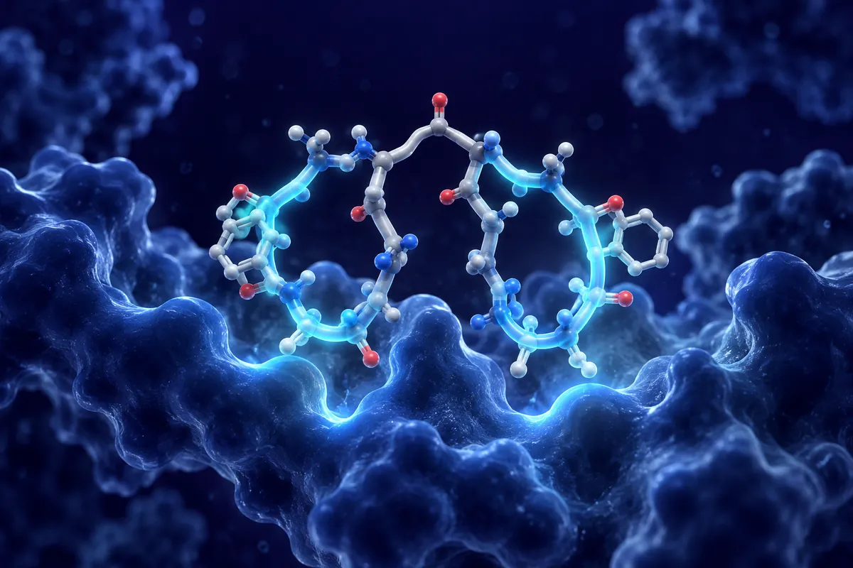
Mitochondria produce much of a cell’s ATP, but their role extends beyond energy production. Their membrane structure, lipid composition, and protein organization are also subjects of laboratory research. Molecular tools help researchers examine how these features relate to one another.
SS-31, also known as elamipretide, is a tetrapeptide studied for its association with the inner mitochondrial membrane. Its four-residue structure and interactions with membrane lipids are the focus of this article.
All content presented here is for research and educational purposes only.
What Is SS-31 (Elamipretide)?
SS-31 belongs to a family of Szeto-Schiller (SS) peptides developed by researchers Hazel Szeto and Peter Schiller. Its structural designation is D-Arg-2’6′-dimethylTyr-Lys-Phe-NH2. The sequence contains an N-terminal D-arginine residue, a modified dimethyltyrosine, lysine, and phenylalanine with a C-terminal amide. The dimethyl substitution contributes to interactions with anionic phospholipids and may affect susceptibility to enzymatic degradation.
In experimental systems, SS-31 has been reported to accumulate in the inner mitochondrial membrane (IMM) at concentrations higher than those in the surrounding cytosol. Its cationic and aromatic residues contribute to interactions with cardiolipin, a phospholipid enriched in the IMM.
All research applications reviewed here remain within the context of scientific investigation.
Cardiolipin’s structure helps explain its role in mitochondrial membranes. Unlike most membrane phospholipids, it has four fatty acid chains and two phosphate groups linked through a central glycerol. It is present at high concentrations in the IMM, where its molecular shape contributes to membrane organization.
Cardiolipin and Mitochondrial Architecture
Cardiolipin plays an almost architectural role in the IMM. Cardiolipin is the molecular glue that holds these arrangements together.
Cardiolipin contributes to IMM curvature at the cristae, the folds where ATP synthase and electron transport chain (ETC) complexes are concentrated. Oxidation or depletion of cardiolipin can alter membrane organization, respiratory supercomplex assembly, and the electrochemical gradient used for ATP synthesis.
This is why cardiolipin damage is so consequential. It’s not just about losing a single phospholipid; it’s about destabilizing the entire architecture that efficient oxidative phosphorylation depends on.
How SS-31 Binds Cardiolipin
SS-31’s interaction with cardiolipin is electrostatic and hydrophobic in character. The D-Arg residue carries a positive charge that is drawn to the anionic phosphate groups of cardiolipin, while the aromatic residues (dimethylTyr and Phe) insert into the hydrophobic core of the membrane. Taken together, this gives SS-31 a snug, high-affinity interaction with cardiolipin-rich regions of the IMM.
The molecular relationships of interest include cardiolipin binding and cytochrome c association. Cytochrome c associates with cardiolipin in the IMM, and cardiolipin oxidation can alter that interaction.
Researchers have characterized effects at multiple nodes of mitochondrial function.
Experimental studies of SS-31 have measured ATP synthesis and ETC supercomplex organization.
The proton gradient across the IMM supplies the electrochemical energy used by ATP synthase (Complex V). Studies have also examined possible interactions between SS-31 and ATP synthase, although the details remain under investigation.
Reactive Oxygen Species Modulation
Mitochondria generate reactive oxygen species (ROS), including superoxide and hydrogen peroxide. These molecules participate in signaling and, at higher levels, can oxidize proteins, lipids, and DNA. Cardiolipin is studied in this context because of its proximity to Complexes I and III, two sites of mitochondrial ROS generation.
Cardiac Research Models
Cardiac muscle cells have high mitochondrial content and a continuous demand for ATP. They provide a setting for studying oxidative phosphorylation, membrane organization, and mitochondrial respiration.
Ischemia-Reperfusion Injury Models
In preclinical ischemia-reperfusion (I/R) models, blood flow is interrupted and then restored. SS-31 research in these models has examined mitochondrial function and cellular responses to the resulting changes in oxygen supply.
Findings in these models need to be read alongside the experimental conditions, comparison group, and specific mitochondrial or cellular endpoint measured.
Mitochondrial Dysfunction Models
Mitochondrial dysfunction models vary in their experimental conditions. Differences in tissue preparation and measurement methods matter when comparing findings.
Open questions include how experimental conditions affect mitochondrial measurements and which assay endpoints capture distinct aspects of membrane function.
Other Tissue Research Models
The discussion also extends to skeletal muscle, kidney tissue, and neuronal models.
Skeletal muscle models allow researchers to measure respiratory capacity, ROS production, and ATP synthesis. Studies of aging muscle examine how these mitochondrial measurements vary with age and experimental conditions.
This remains an active area for preclinical model refinement and mechanistic research.
Renal and Neurological Models
Kidney tissue has a high mitochondrial density. Preclinical SS-31 research has included cisplatin-induced and ischemic kidney injury models.
Neurons rely on mitochondrial metabolism. In neuronal research models, mitochondrial respiration and membrane organization can be examined as separate experimental endpoints.
SS-31 vs. Other Mitochondrial Peptides
SS-31 doesn’t exist in isolation. The SS peptide family includes SS-02 and SS-20, and the broader field of mitochondria-targeting compounds includes MitoQ (mitoquinone), SKQ1, and various other antioxidant conjugates. How does SS-31 compare?
SS-02 has μ-opioid receptor activity in addition to its mitochondrial interactions.
Experimental Properties and Assay Observations
SS-31 is water-soluble and has a molecular weight of approximately 640 Da. Preclinical studies have examined its distribution and mitochondrial accumulation over time.
Cell-based studies have examined SS-31 at nanomolar to low micromolar concentrations. The assay system, exposure time, and measured endpoint affect how those concentrations are interpreted. The N-terminal D-arginine residue is also relevant to studies of proteolytic stability.
Standard peptide handling protocols apply.
Future Research Directions
SS-31 research focuses on the relationship between peptide-lipid interactions and mitochondrial measurements. A central question is how cardiolipin association relates to membrane organization and bioenergetic assays.
Future experiments could examine the relationship between cardiolipin organization, NAD+ metabolism, PGC-1α signaling, and mitochondrial biogenesis. Separate measurements would be needed to distinguish these processes.
Further preclinical work could clarify how model conditions, membrane composition, and assay choice affect observations.
Conclusion
SS-31 research spans mitochondrial membranes and several tissue models. Comparing findings requires attention to the experimental system, the conditions tested, and the endpoint measured.
For researchers working in these areas, the mechanistic clarity of SS-31’s action is a distinct scientific asset.
All information presented in this article is for research and educational purposes only. SS-31 (elamipretide) is not approved for human use and is intended exclusively for laboratory and preclinical research.
This educational material discusses laboratory research concepts only. Loti Labs products are intended strictly for research and development use and are not for human consumption.