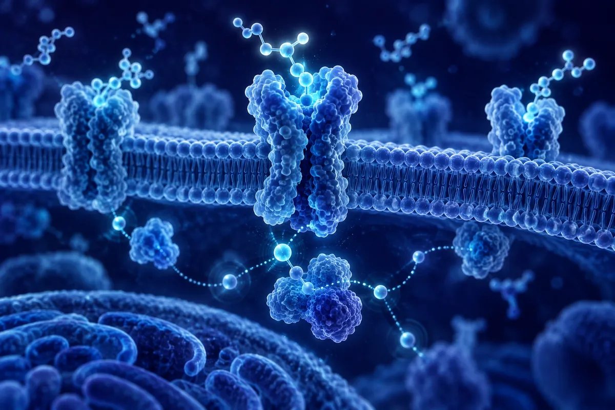
Messenger RNA delivery has a charge problem. RNA is a polyanion, dense with phosphate groups, and getting it across a lipid bilayer without shredding it in the endosome has driven two decades of carrier chemistry. Cationic lipid nanoparticles solved the packaging problem well enough to reach global distribution, but their positively charged components are not inert bystanders. Ionizable lipids and polycationic peptides can perturb membranes, activate innate immune sensors in cultured macrophages, and leave residual cationic material that complicates long-term storage stability. A newer line of peptide engineering asks a different question: can a carrier condense mRNA without relying on a net positive charge at all?
The Case Against Permanent Cationic Charge
Polycationic peptides such as polylysine and protamine derivatives have been used for nucleic acid condensation since before lipofection existed. They work by brute electrostatics : enough positive charge overwhelms the phosphate backbone and collapses RNA into a compact particle. The trouble is that the same charge density that drives condensation also drives nonspecific binding to serum proteins, red blood cell membranes, and off-target cell surfaces in culture. In hemolysis assays run on isolated erythrocytes, strongly cationic peptides above a certain charge density reliably lyse membranes in a concentration-dependent manner. That is a signal, not a footnote : it tells the chemist that charge magnitude and biological tolerability are pulling in opposite directions.
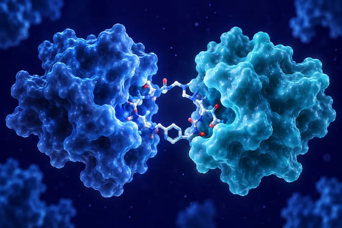
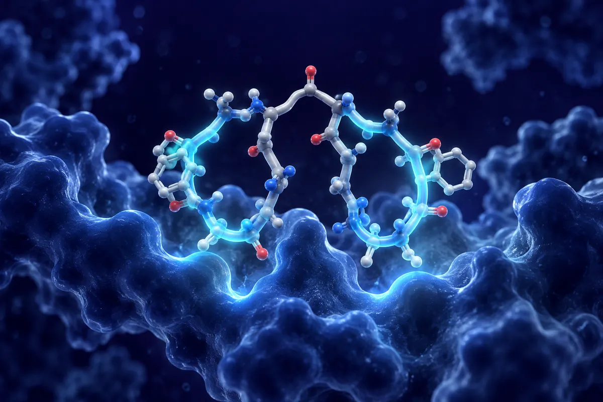
Researchers working on self-assembling peptide systems have tried to decouple those two variables. What if condensation came from hydrogen bonding and hydrophobic packing instead of charge? A peptide sequence built around alternating hydrophobic and polar residues : the classic beta-sheet-forming motif seen in sequences like Ac-(RADA)4-CONH2 and its neutral analogs : can self-assemble into nanofibers and nanotubes in aqueous buffer without carrying a large net charge. When these assemblies are mixed with mRNA in vitro, the RNA appears to template along the fiber surface, guided by short-range hydrogen bonding between backbone amides and possibly by hydrophobic exclusion of water rather than by ionic attraction.
Nanotube Geometry and mRNA Templating
The nanotube architecture matters mechanically as well as chemically. Beta-sheet peptides that assemble into hollow or ribbon-like nanotubes present a curved, repeating surface with a periodicity spanning a few nanometers : roughly matched to the helical pitch of duplex RNA secondary structure. Cryo-electron microscopy and small-angle X-ray scattering of related peptide nanotube systems have shown that this periodicity is not accidental; it emerges from the same backbone hydrogen-bonding register that stabilizes the beta-sheet itself. Does that geometric match actually improve loading efficiency for a full-length mRNA transcript, or is it more relevant for short oligonucleotides? That remains an open question, and the honest answer from the current literature is that transcript-length dependence has not been systematically mapped.
What has been shown in cell-free binding assays is that these neutral or near-neutral peptide assemblies can protect associated RNA from ribonuclease digestion for longer than free RNA alone, likely through a combination of steric shielding and reduced water accessibility at the fiber surface. That protective effect is measured with gel electrophoresis and fluorescence-quenching assays, not with any claim about biodistribution in an intact organism : the RNase protection data live entirely at the level of biochemistry in a tube.
Toxicity Signals in Cultured Cell Models
Any nucleic acid carrier eventually has to clear a cytotoxicity bar before it is taken seriously as a delivery platform. Standard practice uses immortalized cell lines : HEK293, HeLa, or relevant primary cell isolates : assayed with lactate dehydrogenase release or MTT/MTS metabolic assays across a dose-dependent concentration range. Charge-neutral peptide assemblies have generally shown a wider therapeutic index in these assays compared with polycationic controls at equivalent RNA-binding capacity, largely because they don’t trigger the same membrane-destabilizing contact that cationic polymers do. That said, “wider” is a comparative statement bounded to the specific cell lines and concentration ranges tested; it is not a blanket safety claim, and different peptide sequences within the same neutral-assembly family can behave quite differently depending on hydrophobic residue placement.
Inflammatory signaling is the other axis worth watching. Cationic lipid components are known activators of TLR-adjacent pathways in murine bone-marrow-derived macrophage cultures, producing measurable cytokine induction before any RNA cargo is even added. Neutral peptide nanotubes show a lower baseline activation signal in comparable macrophage assays, though the mechanistic reason : charge, particle size, or backbone presentation : isn’t fully disentangled.
The Endosomal Escape Bottleneck
Getting mRNA into a cell is only half the job; getting it out of the endosome before lysosomal degradation is the step that kills most delivery efficiency, regardless of carrier chemistry. Cationic lipid nanoparticles rely heavily on the “proton sponge” mechanism and pH-triggered ionizable lipid protonation to destabilize the endosomal membrane. A charge-neutral peptide carrier doesn’t have that same pH-switch built in by default, so escape has to come from somewhere else : often engineered fusogenic or membrane-interacting peptide segments appended to the neutral scaffold, sometimes borrowed from viral fusion peptide motifs studied in influenza hemagglutinin or vesicular stomatitis virus glycoprotein.
Fluorescent reporter assays using pH-sensitive dyes or split-GFP endosomal escape constructs in cultured cells are the standard readout here, and escape efficiency varies enormously depending on how the fusogenic segment is grafted onto the nanotube-forming sequence. Getting that balance right : assembly stability on one end, membrane disruption on the other : is the central design tension in this subfield.
Where the Research Stands
Charge-neutral self-assembling peptides represent a genuinely different design philosophy for nucleic acid carriers, built on hydrogen bonding and hydrophobic packing rather than electrostatics. The in vitro and cell-culture data so far are encouraging on the toxicity and RNase-protection fronts, but transcript-length scalability, endosomal escape efficiency, and any behavior in a living organism remain active research questions rather than settled findings. For laboratory groups working at the interface of peptide chemistry and nucleic acid delivery, that gap is exactly where the interesting work is happening now.
Disclaimer: This content is intended for research purposes only and is not meant to constitute medical advice.
This educational material discusses laboratory research concepts only. Loti Labs products are intended strictly for research and development use and are not for human consumption.