The Hunger Hormone’s Deeper Story
Ghrelin occupies a distinctive place in peptide biology. Discovered in 1999 by Masayasu Kojima and colleagues at Kurume University, it was identified through reverse pharmacology: researchers found the receptor first (growth hormone secretagogue receptor type 1a, GHS-R1a), then searched for its endogenous ligand. What they found was a 28-amino-acid peptide produced primarily by endocrine cells in the gastric fundus, bearing an octanoyl modification on its third serine residue.
That fatty acid modification matters for receptor activation. The enzyme ghrelin O-acyltransferase (GOAT) attaches the octanoyl group to the peptide’s third serine residue. This modification is required for ghrelin activation of GHS-R1a, making acylation status an important variable in receptor assays.
GHS-R1a: A Receptor with Constitutive Activity
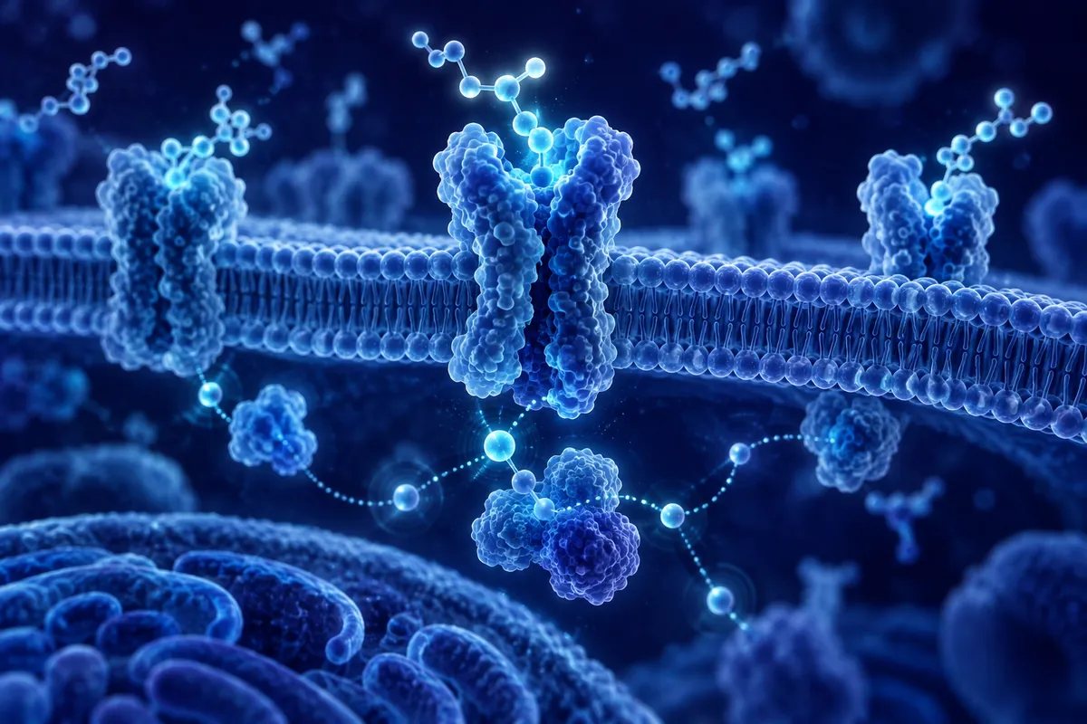
GHS-R1a is remarkable in its own right. Unlike most G-protein-coupled receptors, which remain silent until a ligand arrives, GHS-R1a displays approximately 50% constitutive activity. In the absence of any ghrelin, the receptor signals at half-maximal capacity. Ghrelin binding pushes activity higher; inverse agonists push it lower.
This baseline signaling has significant research implications. Researchers designing experiments with GHS-R1a must account for this constitutive signaling, as receptor deletion produces effects that exceed what ghrelin removal alone would predict.
In isolated pituitary cell assays, adding ghrelin and GHRH together produced GH release greater than the sum of the responses to either peptide alone. The synergy involved distinct but converging intracellular pathways: GHRH signaling through cAMP/PKA and ghrelin signaling through IP3/DAG/PKC. This convergence amplified calcium influx beyond what either pathway generated independently in those assays.
Endogenous ghrelin concentrations vary with feeding state. Levels rise before meals and fall after eating. Sampling time relative to feeding is therefore an important variable when measuring circulating ghrelin.
GHS-R1a signaling is studied in NPY/AgRP neurons of the hypothalamic arcuate nucleus. Vagal afferents connecting the stomach with the nucleus tractus solitarius in the brainstem are another focus of ghrelin signaling research.
These observations describe the experimental systems studied and should not be assumed to hold across species.
Reward, Motivation, and Dopamine
In progressive ratio experiments, rodents exposed to ghrelin pressed levers more times for a food pellet than controls. Both groups were food-deprived to the same extent. These experiments measured operant responding in a rodent model.
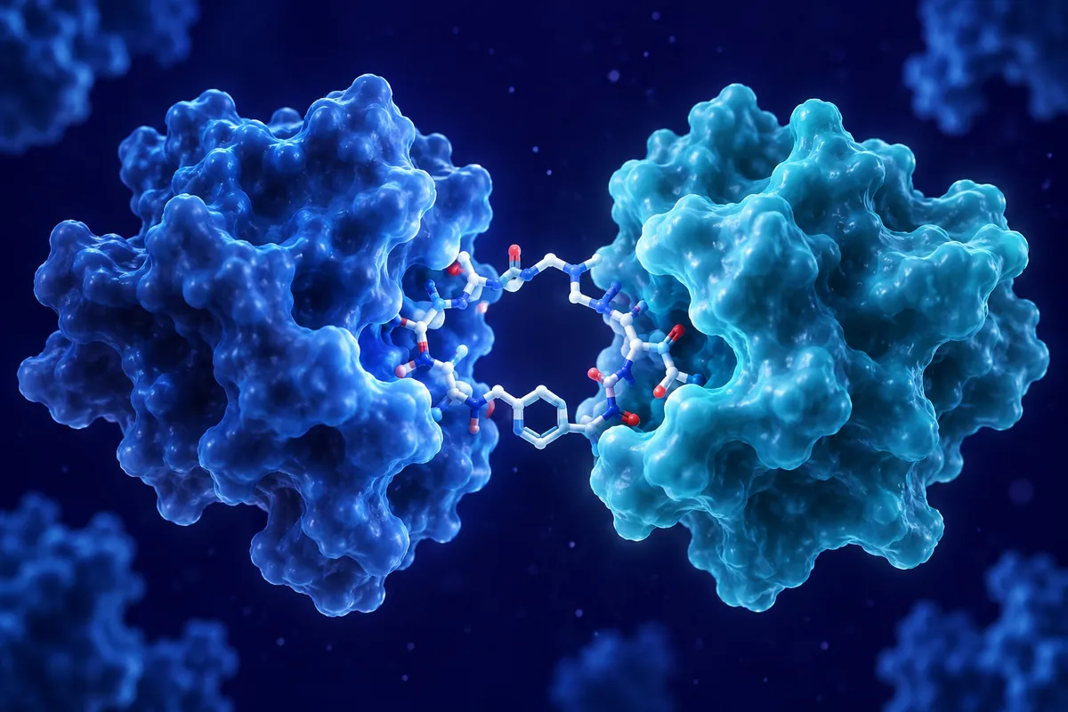
GHS-R1a can form heterodimers with dopamine D1 and D2 receptors, altering their signaling properties. These receptor-receptor interactions are studied to distinguish heterodimer signaling from responses to direct dopamine receptor agonists.
Des-Acyl Ghrelin: The Other Form
Approximately 80 to 90% of circulating ghrelin lacks the octanoyl modification and is designated des-acyl ghrelin.
The acyl/des-acyl ratio therefore matters. Changes in GOAT expression, dietary fatty acid availability (octanoic acid is a medium-chain fatty acid found in coconut and palm kernel oil), and degradation kinetics all influence this ratio. Researchers measuring “total ghrelin” without distinguishing acylation status may miss functionally relevant shifts in the balance between these two forms.
Current Research Directions
Current research examines GHS-R1a signaling in reward circuits and age-related variation in ghrelin pulsatility. Questions include how receptor interactions shape signaling and how ghrelin and GH secretion patterns vary across experimental models.
Disclaimer: This content is intended for research purposes only and is not meant to constitute medical advice.
This educational material discusses laboratory research concepts only. Loti Labs products are intended strictly for research and development use and are not for human consumption.