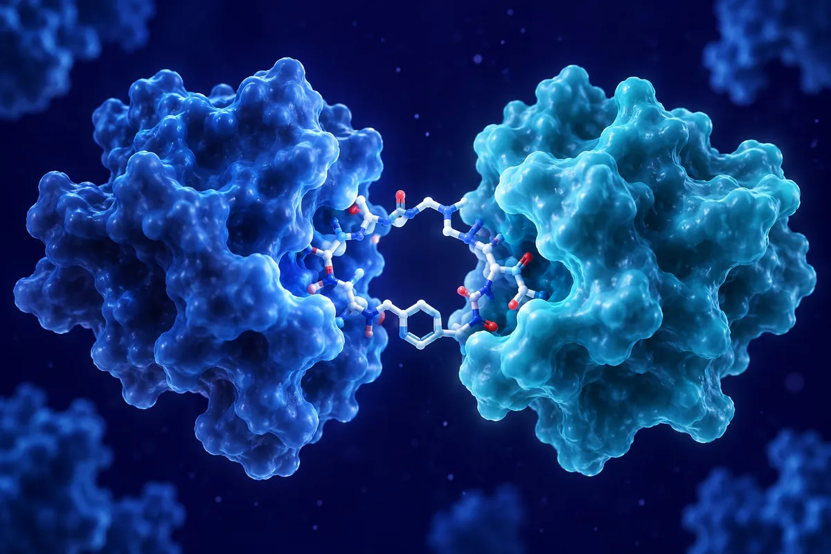
The extracellular matrix isn’t just scaffolding. When enzymes chew up collagen, some of the fragments turn out to be surprisingly active signaling molecules, and a few of them push back hard against new blood vessel growth.
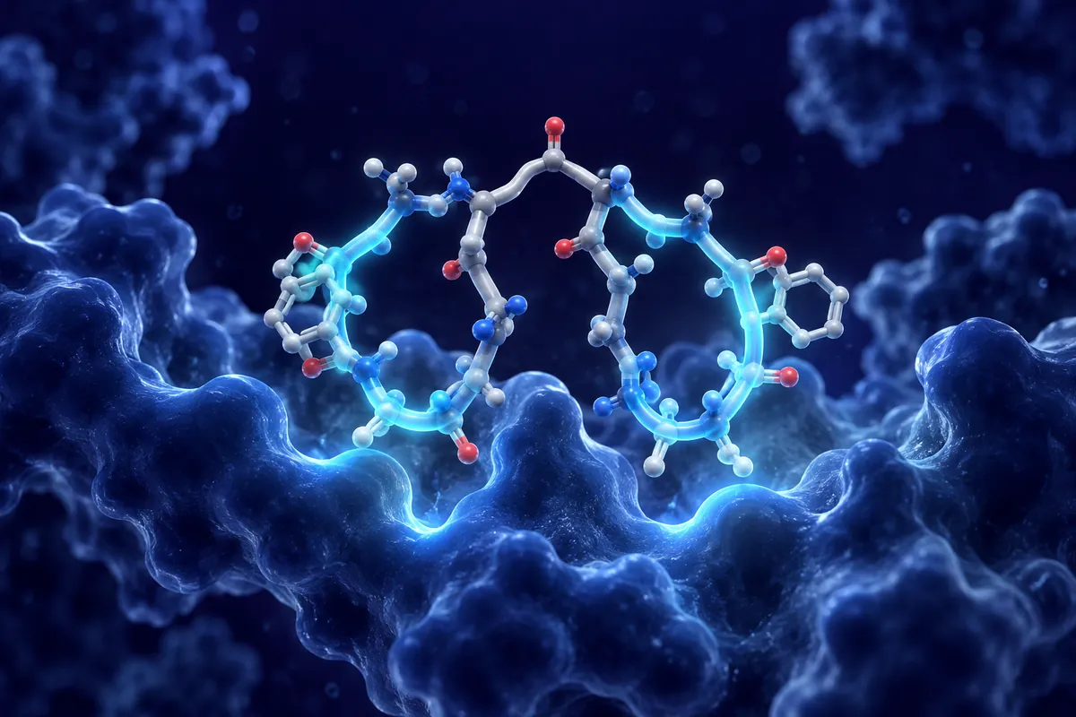
EB-203 is one of the newer entries in that story. A September 2026 paper in Scientific Reports described this collagen-derived peptide in mouse models of choroidal neovascularization (CNV), the abnormal vessel growth beneath the retina that researchers use to study wet age-related macular degeneration biology. Here’s what that work fits into, and what to watch for.
Matrix Fragments That Block Angiogenesis
Back in the 1990s, Judah Folkman’s lab found that endostatin, a C-terminal fragment of collagen XVIII, inhibited endothelial cell growth. It was a big moment. Suddenly the matrix looked like a source of built-in angiogenesis brakes.
Others followed. Type IV collagen, the main collagen of basement membranes, yields several fragments with antiangiogenic activity, including tumstatin, arresten and canstatin, each derived from a different alpha chain’s NC1 domain. Smaller peptides cut down from these domains often keep the activity, which made them attractive for lab work since short peptides are easier to synthesize and modify.
Most of these fragments act through integrins, the receptors that cells use to grip the matrix. Tumstatin, for instance, binds alpha-v beta-3 integrin on endothelial cells. Other fragments influence VEGF receptor signaling or interfere with endothelial migration directly.
Why the eye?
The retina is a favorite testing ground. It’s accessible, vessel growth can be imaged directly, and the laser-induced CNV model in mice is well characterized. A local route of administration (intravitreal delivery in rodents) also keeps systemic exposure low, which simplifies interpretation.
What makes a fragment antiangiogenic while the intact collagen is not? Part of the answer is exposure. Cryptic sites buried inside the folded protein only become visible after proteolysis, so matrix metalloproteinases effectively switch these signals on. It’s a tidy way for tissue to limit vessel growth right where remodeling is happening, and it means the enzymes present in a lesion can shape which fragments appear there.
The Laser-Induced CNV Model
In this model, a laser creates small burns that rupture Bruch’s membrane, the thin matrix layer between the retinal pigment epithelium and the choroid. Over the following week or two, new vessels sprout from the choroid into the damaged area.
Researchers then measure lesion size, often on flat-mounted tissue stained with isolectin B4, or track leakage with fluorescein angiography. Optical coherence tomography adds a cross-sectional view. It’s a fast, reproducible system, although lesion size varies a lot between burns, so you need plenty of them per group.
One honest caveat. Laser CNV is an injury-driven model, and it doesn’t capture the slow, age-related changes that precede vessel growth in natural disease. It tells you whether a compound can limit an acute angiogenic response. That’s useful, but narrower than it sounds.
What the EB-203 Work Reported
According to the Scientific Reports study, EB-203 reduced CNV lesion area in mice compared with control groups. The authors also examined endothelial cell behavior in vitro, looking at proliferation, migration and tube formation, the standard trio of angiogenesis assays. We won’t paraphrase specific percentages here, because the paper itself is the right place for those numbers.
Mechanistic work in studies like this usually checks integrin binding and downstream signaling such as focal adhesion kinase or ERK phosphorylation. Comparing a new peptide with an anti-VEGF reagent in the same model is also common, since VEGF blockade is the reference point everyone knows. Did the peptide add anything beyond VEGF suppression? That’s the question we’d most want answered.
Reading the results critically
Check the controls. A scrambled peptide is better than vehicle alone. Look at how many lesions and animals were counted, and whether the people scoring lesions were blinded. Also notice the timing: giving a compound at the moment of laser injury tests prevention, while giving it later tests regression of established vessels.
Working With Collagen-Derived Peptides
Short matrix peptides can be hydrophobic and prone to aggregation, so solubility testing comes first. Some groups add solubilizing residues or use small amounts of co-solvent. Whatever you choose, keep the vehicle identical across groups.
Stability in the vitreous is another practical issue. Proteases in ocular tissue can shorten a peptide’s half-life considerably, and measuring how long it persists helps explain why an effect appears or fades. Labeled peptide and imaging, or mass spectrometry on tissue extracts, can answer that.
Finally, there’s the integrin question. Many endothelial integrins are also expressed on other retinal cell types, including pigment epithelium and microglia. Off-target activity within the eye is plausible, and histology beyond the vessel stain can reveal it.
Retinal function tests, such as electroretinography in mice, are a sensible add-on. If a peptide shrinks lesions but also dulls photoreceptor responses, that’s worth knowing early.
Conclusion
EB-203 adds to a long line of matrix-derived fragments that slow vessel growth in preclinical models, from endostatin onward. The September 2026 mouse CNV data suggest it limits laser-induced neovascularization, which is a reasonable first step. The more interesting work comes next: defining its receptor targets, testing it against established vessels and comparing it head to head with VEGF blockade. For labs studying retinal angiogenesis or basement membrane biology, it’s a peptide worth following.
Disclaimer: This content is intended for research purposes only and is not meant to constitute medical advice.
This educational material discusses laboratory research concepts only. Loti Labs products are intended strictly for research and development use and are not for human consumption.