Validating peptide purity through High-Performance Liquid Chromatography (HPLC) is the foundational step in interpreting any legitimate Certificate of Analysis (COA). For academic researchers and laboratory managers, mastering the nuances of chromatographic trace data is essential for ensuring experimental reproducibility in preclinical models. This comprehensive guide details the mechanics of chromatographic separation, the critical distinction between total purity and active peptide mass, and the rigorous analytical standards required for modern in vitro investigations.
HPLC peptide testing measures the exact purity of a synthesized peptide batch by separating it from synthesis by-products and quantifying the target compound relative to the total detected sample. The process follows a sequential analytical workflow:
Separates the target peptide from synthesis by-products, deletion sequences, and oxidized variants using a pressurized C18 column.
Measures the UV absorbance of peptide bonds at 214–220 nm, producing a chromatographic trace with area-proportional peaks.
Calculates purity as the percentage of the target peak area against the total integrated area of all detected peptide peaks.
Generates a chromatogram that documents each batch’s purity profile — the primary evidence in every Certificate of Analysis (COA).
High-Performance Liquid Chromatography represents the cornerstone of modern analytical chemistry, specifically engineered to separate, identify, and quantify individual components within a complex chemical mixture. In the context of synthetic peptide research, HPLC is utilized to isolate the target amino acid sequence from synthesis byproducts, truncated sequences, and degradation artifacts. The system operates by forcing a liquid solvent—known as the mobile phase—through a densely packed column containing a solid adsorbent material, referred to as the stationary phase, under extremely high pressures typically ranging from 400 to 600 bar.
The fundamental principle governing this separation technique is differential partitioning. As the dissolved peptide sample is injected into the high-pressure fluid stream, the various molecules within the sample interact with the stationary phase to varying degrees. Molecules that exhibit a stronger affinity for the stationary phase will progress through the column at a slower velocity, whereas molecules with a higher affinity for the mobile phase will elute more rapidly. This differential migration allows an analytical chemist to separate structurally similar compounds that would otherwise be indistinguishable in a bulk assay.
Modern HPLC systems utilized in peptide analysis are highly sophisticated instruments comprising several critical modules: a solvent degasser to prevent bubble formation, precision dual-piston pumps to maintain consistent flow rates, an automated sample injector (autosampler) capable of microliter precision, a temperature-controlled column compartment to ensure thermodynamic stability during separation, and highly sensitive optical detectors. The intricate interplay between these components ensures that the resulting analytical data is both highly reproducible and scientifically rigorous, forming the basis of all legitimate quality control protocols in synthetic peptide manufacturing.
Why Reverse-Phase HPLC (RP-HPLC) Is the Gold Standard
While various modes of chromatography exist, Reverse-Phase HPLC (RP-HPLC) is universally recognized as the gold standard for analyzing synthetic peptides. In traditional “normal-phase” chromatography, the stationary phase is highly polar (e.g., bare silica) and the mobile phase is non-polar. However, peptides are inherently amphipathic molecules containing both hydrophilic and hydrophobic regions, making normal-phase techniques highly inefficient for their separation. Reverse-phase chromatography inverts this paradigm by utilizing a non-polar stationary phase and a polar, aqueous mobile phase.
The stationary phase in RP-HPLC typically consists of silica particles that have been chemically modified with long hydrocarbon chains. The most prevalent configuration for peptide analysis is the C18 column, which employs octadecylsilane (an 18-carbon alkyl chain) bonded to the silica support. This dense, hydrophobic matrix provides an ideal environment for interacting with the hydrophobic side chains of the amino acid residues comprising the peptide. The longer the carbon chain on the stationary phase, the greater the hydrophobic retention, making C18 optimal for resolving closely related peptide sequences that differ by only a single amino acid deletion or substitution.
To elute the tightly bound peptides from the C18 column, analysts employ a mobile phase gradient. Rather than using a single, unchanging solvent composition—a technique known as isocratic elution, which is generally insufficient for complex peptide mixtures—a gradient system dynamically alters the ratio of the solvents over time. The process typically begins with a highly aqueous solvent (such as water) to promote initial binding to the column, followed by a gradual, linear increase in a non-polar organic modifier. As the concentration of the organic solvent increases, it effectively outcompetes the stationary phase for the hydrophobic domains of the peptide, causing the peptide to partition back into the mobile phase and elute from the column into the detector.
Sample Preparation: From Lyophilized Powder to HPLC Injection
Before any chromatographic separation can occur, the lyophilized peptide powder must be prepared into a suitable injection-ready solution. The sample preparation protocol directly affects chromatographic reproducibility and the reliability of the resulting purity data. Standard preparation steps include:
Dissolution: The lyophilized peptide is weighed on an analytical balance and dissolved in a compatible solvent — typically a mixture of acetonitrile and ultrapure water (20:80 v/v) containing 0.1% TFA — at a target concentration of 0.5–1.0 mg/mL for analytical injection.
Filtration: The dissolved sample is passed through a 0.22 μm PVDF or nylon membrane filter to remove any particulate matter that could block the HPLC column frit or cause abnormal baseline spikes.
Degassing: The mobile phase solvents are degassed (via sonication or sparging with helium) to prevent dissolved gas from forming bubbles in the detector flow cell, which cause spurious baseline noise.
Column Equilibration: The C18 column is equilibrated with the starting mobile phase composition (typically 95% aqueous / 5% organic) for at least 10 column volumes prior to injection to establish a stable, reproducible baseline.
Injection Volume: A defined injection volume (typically 5–20 μL) is loaded onto the column via the autosampler, ensuring consistent sample delivery across multiple injections.
Each of these steps is governed by the validated method parameters that the analytical chemist documents in the laboratory’s standard operating procedure (SOP). Deviations from these parameters — such as incomplete dissolution or insufficient column equilibration — directly compromise the reliability of the purity result and represent a methodological risk when evaluating vendor-issued COA data from undisclosed or non-validated procedures.
UHPLC vs. HPLC: The 2026 Standard for Complex Peptides
Ultra-High Performance Liquid Chromatography (UHPLC) represents an evolution of classical HPLC that operates at substantially higher system pressures (up to 15,000–18,000 psi versus HPLC’s typical 5,000–6,000 psi) using sub-2-micron column particles rather than the conventional 3–5 micron particles of standard C18 columns. The result is dramatically improved chromatographic resolution, narrower peak widths, and significantly reduced run times — typically 5–12 minutes for a UHPLC gradient versus 20–30 minutes for a conventional HPLC method. For simple, well-characterized peptides of 5–15 residues, classical RP-HPLC on a Vydac C18 column remains the standard release method. However, for larger, structurally complex peptides — including the next-generation GLP-1 receptor agonists like Retatrutide (a 39-residue triple agonist) and the gonadotropin HCG — UHPLC provides superior resolution of structurally similar deletion sequences and post-translational modification variants that would co-elute under standard gradient conditions. As of 2026, progressive third-party analytical laboratories have adopted UHPLC as the preferred platform for complex peptide QC, with results deliverable in 48–96 hours. Researchers evaluating vendor COAs for higher-molecular-weight research compounds should verify whether the reported purity was generated on a validated UHPLC or conventional HPLC platform — a methodological detail that directly affects the resolving power applied to the impurity profile.
The Anatomy of a Peptide HPLC Chromatogram
Retention Time (Rt) and the X-Axis
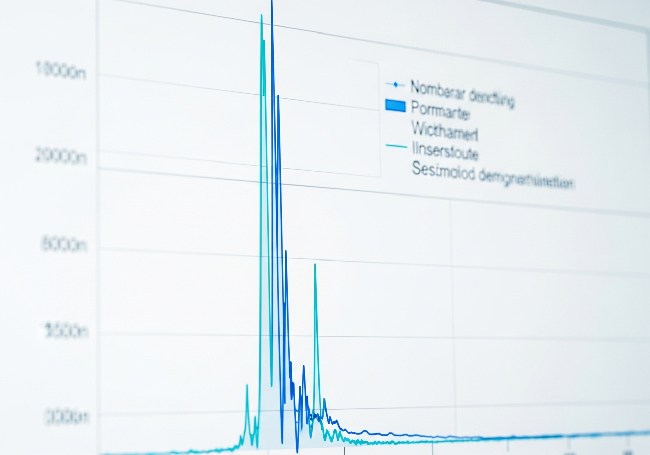
The primary visual output of an HPLC analysis is the chromatogram, a two-dimensional plot that maps the detector signal against time. The horizontal X-axis of this plot represents time, measured in minutes, from the exact moment the sample is injected into the system to the moment the final compound elutes. The specific point on this axis where a particular compound reaches its maximum concentration in the detector is designated as the Retention time (Rt). In a properly calibrated and standardized HPLC method, the Rt is a highly reproducible characteristic of a specific molecule under exact chromatographic conditions.
For synthetic peptides, the retention time is governed by the molecule’s overall hydrophobicity, its three-dimensional conformation in solution, and its molecular weight. Peptides with a higher proportion of hydrophobic amino acids (such as leucine, isoleucine, and valine) will exhibit stronger interactions with the C18 stationary phase, resulting in a longer retention time. Conversely, highly polar or charged peptides will elute earlier in the gradient. Academic researchers scrutinizing a COA must verify that the target peptide’s retention time aligns with expected theoretical models and historical batch data, as unexpected shifts in Rt can indicate structural alterations, incorrect sequence synthesis, or uncalibrated instrumentation.
UV Detection (214-220 nm) and the Y-Axis
The vertical Y-axis of the chromatogram represents the intensity of the signal generated by the molecules as they pass through the detector, typically quantified in milli-Absorbance Units (mAU). For peptide analysis, the universal standard is an Ultraviolet (UV) detector at 214-220 nm. This specific wavelength range is not chosen arbitrarily; it corresponds directly to the maximum absorption frequency of the peptide bond (the amide linkage) that connects individual amino acids.
When the eluting fluid passes through the detector’s flow cell, a UV lamp directs light at the 214-220 nm wavelength through the liquid. Because every peptide bond within the molecule absorbs this specific wavelength of light, the total reduction in light transmission is directly proportional to the concentration of the peptide in the solution, in accordance with the Beer-Lambert Law. This makes UV detection at 214 nm an exceptionally sensitive and universal method for detecting all peptidic material, regardless of the specific amino acid sequence. While some laboratories may simultaneously monitor 254 nm or 280 nm to detect specific aromatic amino acids (like tryptophan or tyrosine), the 214-220 nm trace remains the definitive standard for calculating overall peptide purity.
Baselines, Peaks, and Peak Area (Area%)
A pristine chromatogram begins with a stable Chromatogram baseline, which is the steady-state signal generated by the pure mobile phase flowing through the detector in the absence of any injected sample. When an eluting compound enters the flow cell, it absorbs the UV light, causing the signal to rise sharply above the baseline, reach a maximum vertex (the retention time), and then return to the baseline, thereby forming a two-dimensional peak. The geometric shape, symmetry, and width of this peak provide critical information regarding the efficiency of the chromatographic separation.
The most crucial metric derived from the chromatogram is the Peak Area (Area%). Modern chromatography data systems (CDS) utilize advanced integration algorithms to calculate the exact area under the curve for every peak detected above the baseline noise threshold. The Area% of the primary peak is calculated by dividing its integrated area by the total integrated area of all peaks present in the chromatogram. When a vendor’s COA claims “99% Purity,” they are specifically stating that the main target peak accounts for 99% of the total UV absorbance detected at 214 nm. It is imperative to understand that Area% is a measure of relative chromatographic purity, not an absolute measurement of total peptide mass.
Identifying Peptide Impurities on a Trace
Tailing, Fronting, and Co-elution (Shoulders)
The visual morphology of the peaks on an HPLC trace provides vital diagnostic clues regarding the quality of both the peptide and the analytical method itself. In an ideal theoretical model, an eluting peptide produces a perfectly symmetrical, Gaussian-shaped peak. However, in practical laboratory settings, deviations from this ideal geometry are common and must be critically evaluated. “Tailing” occurs when the right side (the trailing edge) of the peak is elongated and fails to return sharply to the baseline. This phenomenon frequently indicates secondary, unwanted chemical interactions between the peptide and unbonded silanol groups on the silica support matrix, or column degradation.
Conversely, “fronting” presents as an asymmetrical peak with a sloping leading edge and a sharp trailing edge. This is typically a symptom of column overloading, where the concentration of the injected sample exceeds the binding capacity of the stationary phase, causing a portion of the peptide to rush ahead of the main band. The most critical impurity indicator, however, is the presence of “shoulders”—small, secondary peaks that merge into the ascending or descending slope of the main peak. Shoulders indicate co-elution, meaning an impurity possesses a retention time so similar to the target peptide that the current gradient method cannot fully separate them. This is a common hallmark of structurally similar synthesis errors, such as deletion sequences (n-1 peptides) lacking a single amino acid.
Baseline Drift vs. Instrumental Noise
Accurate peak integration relies heavily on the stability of the chromatogram baseline. In RP-HPLC utilizing a mobile phase gradient, the composition of the solvent changes dramatically over the course of the run, typically transitioning from a high concentration of water to a high concentration of Acetonitrile. Acetonitrile is the preferred organic modifier in peptide chromatography due to its low viscosity, excellent UV transparency at 214 nm, and strong eluting power. However, as the ratio of acetonitrile increases, the optical refractive index of the mobile phase changes, which can cause a gradual, upward or downward slope in the detector signal known as baseline drift.
To counteract this phenomenon and maintain sharp peak shapes, analytical methods employ ion-pairing agents, the most standard being Trifluoroacetic acid (TFA). TFA is added to both the aqueous and organic mobile phases (typically at a concentration of 0.1% v/v). It serves a dual purpose: it lowers the pH to fully protonate the peptide’s basic residues, and its hydrophobic trifluoromethyl group pairs with these positive charges to increase the peptide’s affinity for the C18 column. While TFA significantly improves peak resolution and minimizes baseline drift, distinguishing between natural gradient drift and actual instrumental noise (rapid, random fluctuations in the baseline caused by pump cavitation, dirty flow cells, or electronic interference) is a critical skill for evaluating the reliability of a purity report.
Peptide Purity vs. Net Peptide Content (NPC)
Purity (Area%) Explained
One of the most pervasive misunderstandings in preclinical peptide research is the conflation of chromatographic purity with absolute peptide mass. When an HPLC report indicates a purity of 99%, this metric solely reflects the Area% of the main peak relative to other UV-absorbing impurities detected during the chromatographic run. It confirms that out of all the synthesized peptidic material present in the sample, 99% consists of the target sequence, free from significant truncated sequences, oxidation products, or synthesis byproducts.
However, HPLC purity (Area%) is entirely blind to non-UV-absorbing mass. It does not account for the presence of salts, counter-ions, residual solvents, or ambient moisture that may be bound to the peptide matrix. Therefore, a vial containing a highly pure peptide (99% Area%) does not mean that 99% of the physical weight inside that vial is the active peptide molecule. To determine the actual amount of active substance available for an in vitro assay, researchers must understand and calculate the Net Peptide Content (NPC).
Factoring In Counter-ions (TFA/Acetate) and Moisture
During the final stages of synthesis and purification, peptides are subjected to lyophilization, a rigorous freeze-drying process that converts the purified liquid solution into a stable, solid Lyophilized powder. Because peptides are typically purified using RP-HPLC mobile phases containing TFA, the resulting lyophilized powder is not a free-base molecule; it is a peptide salt. The basic amino acid residues within the peptide sequence (such as Arginine, Lysine, and Histidine), as well as the free N-terminus, carry positive charges that stoichiometrically bind with the negatively charged trifluoroacetate counter-ions.
These TFA counter-ions possess significant molecular weight but do not absorb UV light at the same frequencies as peptide bonds, rendering them invisible on the standard HPLC trace. Furthermore, lyophilized peptide salts are highly hygroscopic, readily absorbing ambient moisture from the atmosphere. Depending on the specific amino acid sequence and the lyophilization conditions, a peptide powder may contain anywhere from 10% to 30% of its total weight as bound TFA salts and water. To accurately quantify this non-peptide mass, rigorous analytical laboratories employ Amino acid analysis (AAA) to determine absolute peptide concentration, and perform a Karl Fischer titration to precisely measure the residual water content.
The Peptide Yield Formula
The critical distinction between HPLC Purity and NPC must be mathematically resolved to ensure accurate dosing in laboratory models. While HPLC Purity confirms the quality of the peptide fraction, NPC represents the percentage of the total gross weight that is actually composed of the peptide molecule itself. Most research-grade peptides exhibit an NPC ranging from 70% to 85%, depending heavily on the number of basic residues in the sequence.
To determine the exact amount of active substance available for an experiment, researchers must apply the following Peptide Yield Formula:
Active Peptide Mass = Gross Weight × (NPC% / 100)
Consider a practical worked example: A laboratory purchases a vial labeled as containing 10 mg of a specific peptide. The accompanying COA shows an exceptional HPLC Purity of 99%, but reports a Net Peptide Content (NPC) of 78%. Applying the formula:
Active Peptide Mass = 10 mg × (78 / 100) = 7.8 mg.
In this scenario, the vial contains exactly 10 mg of total lyophilized powder, but only 7.8 mg of that powder is the active peptide sequence; the remaining 2.2 mg consists of invisible TFA counter-ions and bound moisture. If a researcher assumes the vial contains 10 mg of active peptide and reconstitutes it to create a specific molarity for a receptor binding study, their calculations will be off by 22%, severely compromising the reproducibility and validity of the experimental data. This mathematical breakdown underscores why relying solely on a “99% pure” marketing claim is insufficient for rigorous scientific inquiry.
Why HPLC Requires LC-MS for Complete Verification
Separation (HPLC) vs. Molecular Weight Identity (Mass Spec)
While High-Performance Liquid Chromatography is unparalleled in its ability to separate and quantify the relative purity of a sample, it possesses one fundamental limitation: it cannot definitively identify the chemical structure of the peaks it isolates. An HPLC trace only proves that a substance eluted at a specific time; it does not prove what that substance actually is. To achieve definitive structural verification, the separation power of HPLC must be coupled orthogonally with Mass Spectrometry (LC-MS).
LC-MS systems bridge the gap between physical separation and molecular identification. As the separated peptide elutes from the HPLC column, it is directed into the mass spectrometer, where it is subjected to an ionization source—most commonly Electrospray Ionization (ESI). The ESI process imparts multiple electrical charges to the peptide molecules and converts them into a gaseous state. The mass analyzer then separates these ions based on their mass-to-charge ratio (m/z) in a high-vacuum environment. By mapping these mass-to-charge ratios, the mass spectrometer provides a highly precise measurement of the molecule’s exact molecular weight, allowing researchers to confirm that the major peak observed on the HPLC trace is, in fact, the intended amino acid sequence.
Reading the Mass Spectrum Alongside the Chromatogram
Interpreting an LC-MS report requires analyzing the mass spectrum in tandem with the HPLC chromatogram. Because peptides are relatively large molecules, they typically accept multiple protons (H+) during the ESI process. Consequently, the mass spectrum will not display a single peak representing the total molecular weight; instead, it will display a multiply charged envelope—a series of peaks representing the peptide carrying +2, +3, +4, and +5 charges. A skilled analytical chemist utilizes deconvolution software to mathematically resolve these multiple m/z peaks back to the monoisotopic mass of the uncharged molecule.
If the target peptide has a theoretical molecular weight of 2000.0 Daltons, the LC-MS report must display a deconvoluted mass that matches this theoretical value within a very narrow margin of error (typically ± 1.0 Dalton). If the HPLC trace shows 99% purity, but the LC-MS reveals a molecular weight of 1982.0 Daltons, it immediately indicates a critical synthesis failure—specifically, the loss of a water molecule (18 Daltons), likely due to unintended aspartimide formation or dehydration during cleavage. Therefore, a standalone HPLC report without corroborating LC-MS data is analytically incomplete and insufficient for validating research materials.
How to Verify a Vendor’s Certificate of Analysis
Red Flags on an HPLC Report
Evaluating a vendor’s COA requires a highly skeptical, analytical approach. A legitimate HPLC report must contain comprehensive methodological metadata; a simple image of a peak is meaningless without context. Researchers should look for the specific column parameters, such as the use of an industry-standard Vydac C18 column or equivalent, alongside the exact dimensions (e.g., 4.6 x 250 mm) and particle size (e.g., 5 µm). The report must explicitly detail the mobile phase composition (Phase A and Phase B), the gradient slope (e.g., 10% to 60% B over 30 minutes), the flow rate, and the injection volume.
Critical red flags include chromatograms where the Y-axis (mAU) scale is hidden or truncated, which can obscure baseline noise and artificially inflate the visual appearance of purity. Furthermore, the integration table must list all detected peaks, not just the primary peak. If a chromatogram shows visible secondary peaks or shoulders, but the integration table only lists a single row claiming 100% purity, the data has been manually manipulated or the integration threshold has been improperly set. Legitimate testing facilities adhere strictly to USP <621> guidelines regarding system suitability, peak symmetry, and resolution factors, ensuring that the analytical method is genuinely capable of resolving closely related impurities.
Additional COA completeness criteria that distinguish rigorous from superficial quality documentation include:
Endotoxin test results: For peptides intended for cell culture, ex vivo tissue, or in vivo animal model research, endotoxin contamination is a critical confounding variable. A complete COA should include an endotoxin result from a validated Limulus Amebocyte Lysate (LAL) assay, with reported values below the acceptable research threshold (typically <1.0 EU/mg for cell culture grade material). The absence of endotoxin data does not mean the batch is endotoxin-free — it means the parameter was not tested.
Batch number and testing date: Each COA should carry a unique batch or lot identifier that matches the vial label, along with the specific date the analytical testing was performed. A COA lacking a batch number cannot be verified as specific to the received product. A testing date older than 18–24 months should prompt inquiry about storage conditions and compound stability since testing.
Physical appearance: Legitimate COAs include a physical description of the batch — typically “white to off-white lyophilized powder.” Deviation from this description (yellowing, clumping, visible moisture) is an independent quality flag that cannot be detected by HPLC.
Perfectly rounded purity numbers (e.g., exactly 99.00%, 98.00%): Authentic HPLC integrations produce irregular values with decimal variation (e.g., 98.37%, 99.12%). Uniformly rounded values across multiple products from the same vendor are a statistical improbability and a recognized indicator of fabricated data.
Anonymized Blinded Testing and the Golden Batch Problem
One of the most significant analytical integrity risks in the research peptide market is the practice of selective sample submission — vendors submitting a superior-quality “golden batch” for COA generation rather than a representative production sample. The resulting COA accurately reflects the golden batch but does not represent the product researchers actually receive. This practice is not detectable by reviewing the COA alone, because the document itself may be entirely authentic. The defense against golden batch fraud is anonymized independent testing: a researcher orders a production batch from the vendor through a standard purchase, then submits the received vial directly to an ISO 17025–accredited third-party analytical laboratory — without disclosing the vendor’s identity — and compares the independently measured purity and molecular weight against the vendor’s published COA. In 2026, platforms that broker crowd-sourced, blinded testing — where independent researchers pool resources to fund systematic testing of vendor products without vendor knowledge — have become a primary mechanism for exposing calibration errors and systematic over-reporting of purity values. Independent analyses of crowdsourced blinded test results published in 2026 found discrepancy rates exceeding 70% between vendor-published COA purity values and independently measured purity — meaning the majority of vendor COA data did not match the product actually received by researchers. This statistic has substantially accelerated the adoption of mandatory independent verification as a research best practice. A further dimension of this quality risk is the distinction between label claim and batch-specific yield: a vendor’s label claim states the nominal product specification, while the actual measured quantity in each individual vial — determined by rigorous amino acid analysis (AAA) of the specific received lot — constitutes the true batch claim. Vendors who provide only catalog-level COA documentation (a single test applying to all subsequent production lots) rather than batch-by-batch release testing expose researchers to undocumented inter-lot variability. ISO 17025 accreditation is the international standard (from the International Organization for Standardization) governing the technical competence and management requirements of analytical testing laboratories. When a COA names an ISO 17025–accredited laboratory as the testing facility, the certification provides independent assurance that the laboratory’s methods are validated, instruments are calibrated, and results are reproducible.
The 2026 Batch Testing Standard
The landscape of research peptide procurement has evolved dramatically, driven by an increased demand for analytical transparency and regulatory scrutiny. Following the voluntary closure of Peptide Sciences in early 2026 and prior enforcement actions against Amino Asylum, the industry has undergone a significant paradigm shift. The historical practice of providing “catalog-level” or “sample-level” testing—where a vendor tests a single vial and applies that COA to years of subsequent inventory—is now universally recognized as unacceptable.
Today, third-party, batch-by-batch HPLC and LC-MS testing is the de facto baseline standard for legitimate research peptide vendors. The modern benchmark requires a minimum of ≥98% RP-HPLC purity, alongside confirmed molecular weight identity by LC-MS, conducted for every individual synthesis batch prior to release. Furthermore, static PDF documents are no longer sufficient due to the ease of digital forgery. QR-linked COA hubs integrated with SHA-256 blockchain timestamping have emerged as the 2026 transparency standard. A legitimate vendor’s COA must link directly to a verifiable, tamper-evident record hosted independently by the analytical laboratory, ensuring that the data presented matches the exact batch of the reagent received by the researcher.
Research Use Only (RUO)
All peptide compounds, testing methodologies, and analytical data discussed in this guide are referenced exclusively in the context of in vitro laboratory research and preclinical animal model investigation. None of the compounds, protocols, or analytical standards described herein are approved for human use, clinical application, diagnostic purposes, or therapeutic administration. This content is intended for qualified researchers operating within institutional review and applicable regulatory frameworks.
Frequently Asked Questions
What is HPLC testing for peptides?
High-Performance Liquid Chromatography (HPLC) is an analytical technique used to separate, identify, and quantify the individual components within a synthesized peptide mixture. By pumping a liquid sample through a densely packed column under high pressure, the system separates the target peptide sequence from synthesis errors, truncated byproducts, and degradation artifacts based on their unique chemical interactions with the column matrix.
How do you read a peptide HPLC chromatogram?
Reading a chromatogram involves analyzing a two-dimensional plot where the X-axis represents time (retention time) and the Y-axis represents UV absorbance intensity. The primary target peptide should appear as the dominant, symmetrical peak on the trace. The integration table below the plot calculates the Area% of this peak relative to all other detected peaks, providing the relative chromatographic purity of the sample.
What is the difference between peptide purity and net peptide content (NPC)?
Peptide purity (Area%) measures the percentage of the target peptide relative to other UV-absorbing peptidic impurities in the sample. Net Peptide Content (NPC), however, measures the actual mass percentage of the active peptide relative to the total gross weight of the lyophilized powder, factoring in non-UV-absorbing mass like bound TFA counter-ions and ambient moisture. A highly pure peptide can still have a low NPC.
Why is reverse-phase HPLC used for peptides instead of other HPLC modes?
Reverse-phase HPLC is the preferred mode because peptides are amphipathic molecules with significant hydrophobic regions. A non-polar stationary phase (like a C18 column) paired with a polar, aqueous mobile phase gradient perfectly exploits these hydrophobic interactions, allowing for the precise separation of structurally similar peptide sequences that normal-phase chromatography cannot resolve.
What does 98% peptide purity actually mean?
A claim of 98% peptide purity means that out of all the UV-absorbing chemical entities detected during the HPLC run at a specific wavelength (typically 214 nm), 98% of the total integrated peak area belongs to the target peptide sequence. It indicates a highly successful synthesis with minimal peptidic byproducts, but it does not represent the absolute weight of the active molecule in the vial.
What is a C18 column and why is it used in peptide HPLC?
A C18 column is a specific type of HPLC stationary phase composed of silica particles bonded with octadecylsilane, an 18-carbon alkyl chain. This creates a dense, highly hydrophobic surface. It is the standard for peptide analysis because the long carbon chains provide optimal retention and resolution for the hydrophobic side chains of amino acids, allowing for the separation of complex peptide mixtures.
What is retention time in HPLC and how does it identify a compound?
Retention time is the exact duration, measured in minutes, that it takes for a specific molecule to travel from the point of injection through the column and into the detector. While it does not provide definitive structural identification on its own, a consistent retention time under strictly controlled chromatographic conditions is a highly reproducible characteristic used to confirm the presence of a known target peptide.
How does UV detection work in peptide HPLC?
UV detection in peptide HPLC typically operates at 214-220 nm, the specific wavelength range where the amide bonds connecting amino acids strongly absorb ultraviolet light. As the peptide elutes through the detector’s flow cell, it absorbs the UV light emitted by the lamp. The resulting decrease in light transmission is measured and converted into the signal peaks seen on the chromatogram.
What are peptide impurities and how do they appear on a chromatogram trace?
Peptide impurities are predominantly synthesis errors, such as missing amino acids (deletion sequences), incomplete deprotection byproducts, or degradation artifacts like oxidation. On a chromatogram, these impurities appear as secondary, smaller peaks. If an impurity is structurally very similar to the target peptide, it may co-elute, appearing as an asymmetrical “shoulder” merged onto the main peak.
Why must HPLC data always be paired with LC-MS confirmation?
HPLC is an exceptional tool for physical separation and determining relative purity, but it cannot definitively identify the molecular structure of the peaks it isolates. Mass Spectrometry (LC-MS) is required to measure the exact mass-to-charge ratio of the eluted molecule, confirming that the dominant peak on the HPLC trace actually possesses the correct molecular weight of the intended peptide sequence.
What is baseline drift in HPLC and does it affect reported purity?
Baseline drift is the gradual upward or downward shift of the chromatogram’s baseline signal during a run, typically caused by the changing optical properties of the mobile phase as the gradient increases the ratio of organic solvent. While normal, excessive drift can complicate peak integration. Analytical chemists use ion-pairing agents like TFA and advanced integration software to correct for drift and ensure accurate purity calculations.
What should researchers look for when evaluating a vendor’s HPLC COA?
Researchers should demand batch-specific testing, not catalog-level generalizations. A legitimate COA must include full methodological metadata (column type, gradient, flow rate), a clearly labeled Y-axis scale, an unedited integration table showing all detected peaks, and paired LC-MS data confirming molecular weight. In modern practice, the COA should feature a verifiable, tamper-evident link hosted directly by the third-party analytical laboratory.
How much does independent HPLC peptide testing cost?
Independent third-party HPLC purity testing for a single peptide sample typically costs between $150 and $250 per sample at accredited analytical chemistry laboratories, depending on the complexity of the method and the laboratory’s turnaround tier. Bundled HPLC + LC-MS analysis — the recommended dual-verification standard — generally ranges from $200 to $400 per batch. Researchers who wish to independently verify a vendor’s COA by submitting a blind sample to a separate laboratory (a practice known as brokered blind testing) should budget at this range per compound tested.
How long does peptide HPLC testing take to get results?
Most accredited analytical laboratories return standard HPLC purity results within 4 to 5 business days from sample receipt. Expedited 48-hour turnaround options are available at most major peptide analytical service providers at an additional premium. UHPLC methods, due to shorter run times of 5–12 minutes versus 20–30 minutes for conventional HPLC, can accelerate the analytical throughput within the laboratory — though sample preparation, column equilibration, instrument verification, and report generation time are shared overhead for both platforms and typically represent the majority of total turnaround time.
What is the difference between HPLC and UHPLC for peptide testing?
Ultra-High Performance Liquid Chromatography (UHPLC) uses smaller column particles (sub-2 μm versus conventional 3–5 μm) and higher operating pressures (up to 18,000 psi) to achieve superior chromatographic resolution and faster run times compared to classical HPLC. For standard research peptides of 5–20 residues, classical RP-HPLC provides adequate resolution and remains the most widely validated release method. For larger, structurally complex peptides — such as Retatrutide (39 residues), GLP-1 analogues, or gonadotropins — UHPLC offers meaningfully better separation of closely related deletion sequences and structural variants that would co-elute on a conventional gradient. Researchers reviewing COAs for high-molecular-weight compounds should note which platform generated the data, as UHPLC-based purity values are generally more discriminating and conservative than classical HPLC values for complex sequences.
Does HPLC prove the identity of a research peptide?
No. HPLC testing confirms the purity percentage of the detected material — the fraction attributable to the target compound relative to all detected species — but it cannot confirm molecular identity. Two peptides with different sequences but similar hydrophobicity profiles can co-elute at nearly identical retention times on a C18 column. Confirming that the compound is structurally the correct peptide sequence requires pairing HPLC purity data with Liquid Chromatography-Mass Spectrometry (LC-MS), which verifies molecular weight against the theoretical value for the target sequence.
What are the main red flags on a peptide COA?
Critical red flags on a research peptide COA include: perfectly rounded purity values (e.g., exactly 99.00% or 98.00%) across multiple products — authentic HPLC integration produces irregular decimal values; identical or recycled chromatogram images applied to different products; no unique batch number matching the vial label; missing minor-peak integration table; undisclosed method parameters (column type, mobile phase, gradient, flow rate); and testing performed by the vendor’s own in-house laboratory rather than a named, ISO 17025–accredited third-party facility.
What is a good HPLC purity percentage for research peptides?
For in vitro research applications requiring experimental consistency and reproducible dose-response relationships, an HPLC purity of ≥98% is the baseline standard in 2026. Peptides with ≥95% purity may be acceptable for exploratory screening applications where minor impurity fractions are unlikely to confound primary endpoints. For cell culture, receptor binding assays, or animal model dose-response studies where impurity contributions could introduce confounding variables, ≥98% purity verified by independent third-party testing is the appropriate specification.
How do I send a peptide sample for independent HPLC testing?
For standard lyophilized peptide samples, most ISO 17025–accredited analytical laboratories require no sample preparation prior to shipment. The intact, sealed vial is shipped directly to the facility using their standard intake form, which captures the sample ID, requested analytical tests (HPLC purity, LC-MS identity, or both), and return address. For blinded independent testing — where the researcher does not disclose the vendor’s identity to prevent preferential handling — a sample alias or internal code is substituted for the product name on the intake form. Results are typically returned by email as a PDF analytical report within 4–5 business days.
Continue Your Research
Explore our complete catalog of premium research compounds.