Peptides That Build Themselves
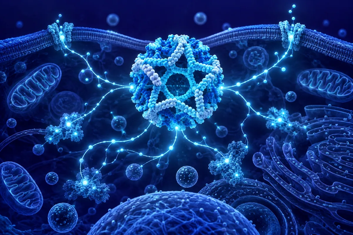
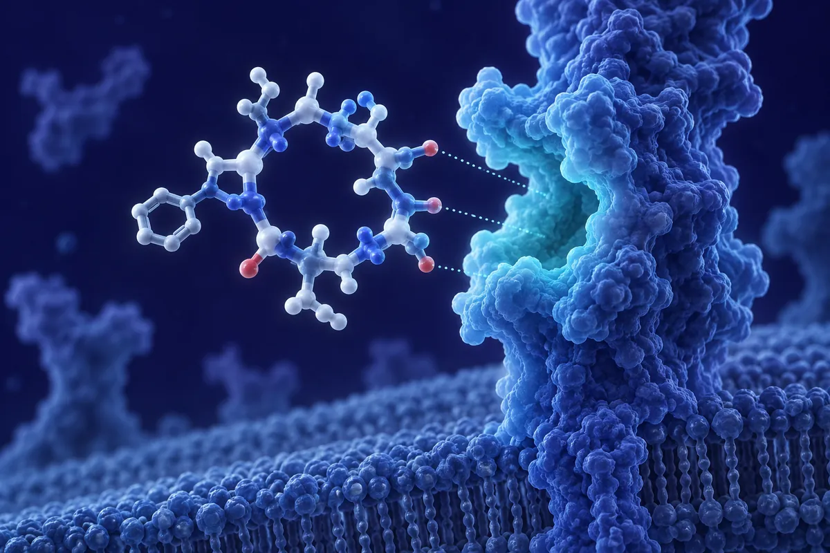
Self-assembling peptides represent one of the most productive intersections of materials science and peptide chemistry. These are short amino acid sequences : often as few as 8-16 residues : that spontaneously organize into ordered nanostructures under physiological conditions. No external template is required. No complex machinery. Just the physics of molecular recognition: hydrogen bonding, hydrophobic interactions, electrostatic complementarity, and aromatic stacking. The peptides do the engineering themselves.
The field traces its modern origins to Shuguang Zhang’s serendipitous discovery at MIT in the early 1990s. While studying a yeast protein involved in DNA repair, Zhang noticed that a segment of the protein : a repeating ionic sequence of alternating hydrophobic and hydrophilic residues : spontaneously formed organized nanofiber networks in aqueous solution. That observation launched a research program that has now generated hundreds of self-assembling peptide designs with applications spanning regenerative research, materials science, and nanomedicine.
Design Principles: Amphipathicity and Complementarity
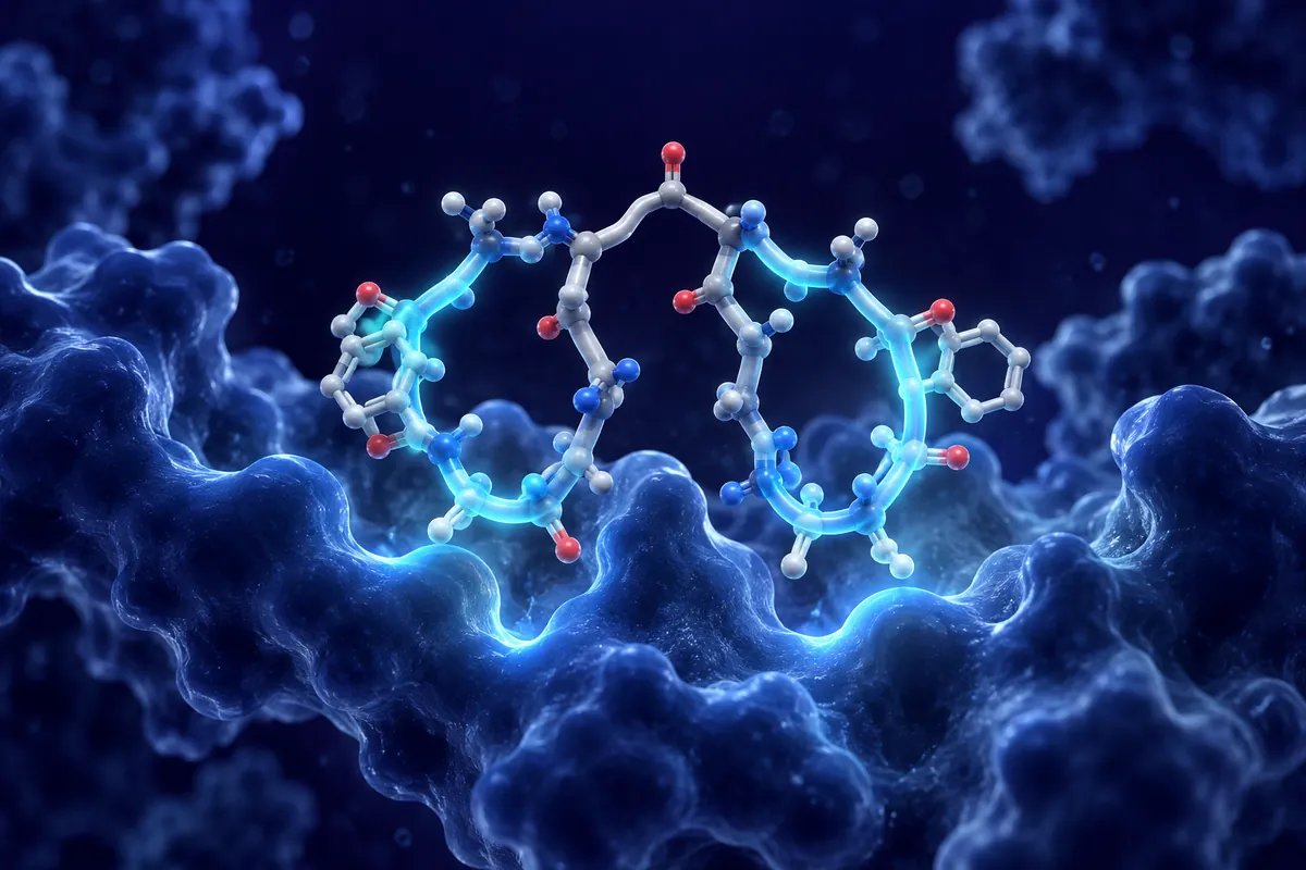
The core design principle is amphipathicity : the arrangement of hydrophobic and hydrophilic residues in a pattern that drives molecular organization. The most studied motif is the ionic complementary peptide, exemplified by the RADA16-I sequence (Ac-RADARADARADARADA-NH₂). Alanine residues form the hydrophobic face; arginine (+) and aspartate (−) residues alternate on the hydrophilic face, creating electrostatic complementarity between adjacent molecules.
In aqueous solution, RADA16-I monomers stack through β-sheet hydrogen bonding along their backbone axis while burying alanine side chains in the interior (hydrophobic collapse) and orienting charged residues toward the solvent. The result: nanofibers approximately 10-20 nm in diameter composed of interdigitated β-sheet bilayers. At sufficient concentration, these nanofibers entangle to form a transparent hydrogel containing over 99% water by mass.
Alternative design motifs include peptide amphiphiles (PA) : molecules combining a hydrophobic alkyl tail with a hydrophilic peptide headgroup : which form cylindrical nanofibers through a combination of hydrophobic collapse and β-sheet formation. Aromatic dipeptides, such as diphenylalanine (FF), self-assemble through π-π stacking into nanotubes and nanofibers with remarkable rigidity. Each architecture offers distinct mechanical properties and functional capabilities.
Hydrogel Formation and Material Properties
The transition from free peptide to hydrogel can be triggered by multiple stimuli: ionic strength changes (adding cell culture media to pure water), pH shifts, temperature adjustments, or even enzymatic catalysis. This stimulus-responsiveness is inherently useful : it allows researchers to prepare peptide solutions that remain liquid until a trigger converts them into structured gels precisely when and where needed.
The mechanical properties of self-assembling peptide hydrogels are tunable across a wide range. By varying peptide concentration, sequence, and assembly conditions, researchers can produce gels with storage moduli spanning from tens of pascals (extremely soft, brain-tissue-like) to thousands of pascals (stiff, approaching cartilage-like). This tunability is critical for tissue engineering research, where the mechanical environment profoundly influences cell behavior, differentiation, and matrix production.
Cell Culture and Three-Dimensional Scaffolds
Self-assembling peptide hydrogels provide a fully synthetic alternative to animal-derived matrices (Matrigel, collagen gels) for three-dimensional cell culture. The advantages are significant: defined composition with no batch-to-batch variability, absence of growth factors or undefined proteins that confound experimental interpretation, and customizable bioactivity through sequence modification.
Cells encapsulated in RADA16-based hydrogels maintain viability, proliferate, and adopt three-dimensional morphologies that differ markedly from flat, two-dimensional culture. Hepatocytes form functional spheroids that produce albumin and urea at rates exceeding standard monolayer culture. Neural progenitor cells extend neurites and form network-like structures. Primary chondrocytes maintain their differentiated phenotype and produce cartilage-specific matrix components : a behavior rapidly lost in conventional 2D culture.
The bioactive version of this approach adds functional motifs to the self-assembling backbone. Appending RGD (cell adhesion), IKVAV (neural adhesion), or GHK (collagen-promoting) sequences to the C-terminus of the assembling peptide incorporates these signals directly into the nanofiber surface. Cells interacting with the gel encounter these motifs in a three-dimensional context that mimics the presentation geometry of native extracellular matrix.
Injectable Applications
Self-assembling peptide solutions are inherently injectable. In liquid form at low ionic strength, they flow through fine-gauge single-use access components without resistance. Upon encountering physiological salt conditions in tissue (approximately 150 mM NaCl), they rapidly transition to a gel state, conforming to the shape of the injection site. This shear-thinning and recovery behavior makes them attractive for minimally invasive delivery research.
In cardiac research models, injectable peptide hydrogels delivered into myocardial tissue formed nanofiber networks that provided mechanical support and recruited endogenous progenitor cells. In spinal cord injury models, self-assembling peptide scaffolds bearing IKVAV sequences promoted axonal regeneration across lesion sites in rodent studies : one of the most demanding regenerative challenges in neuroscience research.
Hemostatic Applications
An unexpected but practical application emerged from the observation that self-assembling peptide solutions can achieve rapid hemostasis : cessation of bleeding : when applied directly to wound surfaces. The peptides assemble into a nanofiber barrier at the tissue interface, physically sealing damaged blood vessels and providing a matrix for platelet adhesion. In preclinical models of liver laceration and femoral artery puncture, RADA16-based solutions stopped bleeding within seconds, outperforming conventional hemostatic agents in time-to-hemostasis comparisons.
Challenges and Future Directions
Self-assembling peptides are not without limitations. Degradation rates in vivo can be unpredictable, influenced by local protease activity that varies between tissues and pathological states. Mechanical strength, while tunable, does not yet match load-bearing tissues like bone or tendon without reinforcement strategies. And the relationship between nanofiber architecture (fiber diameter, pore size, network connectivity) and cellular response is still being mapped systematically.
Yet the trajectory is clear. These peptides offer a bottom-up, molecularly defined approach to constructing three-dimensional research environments with complexity approaching : but never replicating : native tissue architecture. The gap between synthetic scaffold and biological matrix continues to narrow with each generation of designed sequences.
Disclaimer: This content is intended for research purposes only and is not meant to constitute medical advice.
This educational material discusses laboratory research concepts only. Loti Labs products are intended strictly for research and development use and are not for human consumption.