When Peptide Meets Nucleic Acid
Peptide nucleic acids sit at a fascinating intersection of chemistry. They are neither true peptides nor conventional nucleic acids, but a synthetic hybrid that borrows structural logic from both. First described by Peter Nielsen, Michael Egholm, and Ole Buchardt in 1991 at the University of Copenhagen, PNAs replaced the sugar-phosphate backbone of DNA with a polyamide (peptide-like) backbone while retaining the nucleobases : adenine, guanine, cytosine, and thymine : that enable Watson-Crick base pairing.
The result is a molecule that reads genetic sequences with extraordinary specificity but carries none of the negative charge that defines natural nucleic acids. This charge neutrality has profound consequences for binding, stability, and cellular behavior that set PNAs apart from every other class of nucleic acid analog.
Architecture: The N-(2-aminoethyl)glycine Backbone
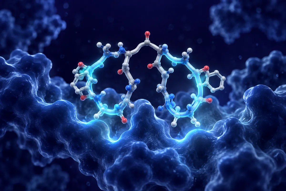
Each PNA monomer consists of a nucleobase attached via a methylene carbonyl linker to an N-(2-aminoethyl)glycine unit. When polymerized, these units form a repeating backbone that is remarkably similar in length and geometry to DNA’s sugar-phosphate chain. The inter-base spacing is virtually identical, allowing PNA strands to hybridize with complementary DNA or RNA sequences through standard base pairing rules.
But the differences matter enormously. DNA’s backbone carries one negative charge per nucleotide from its phosphodiester bonds. PNA’s backbone is electrically neutral. When a PNA strand binds complementary DNA, the absence of charge-charge repulsion between backbones produces a hybrid duplex with higher thermal stability (increased melting temperature, or Tm) than the equivalent DNA:DNA duplex. For a typical 15-mer, the PNA:DNA Tm exceeds the DNA:DNA Tm by approximately 1-1.5°C per base pair : a substantial difference when working with short probe sequences.
Nuclease and Protease Resistance
Here is where PNA’s synthetic origins become a practical advantage. Because the backbone is not a phosphodiester (it lacks the bonds that nucleases cleave), PNAs are completely resistant to DNases, RNases, and restriction endonucleases. Simultaneously, because the backbone is not a standard peptide bond arrangement recognized by proteases, PNAs resist proteolytic degradation as well.
This dual resistance gives PNAs exceptional biostability in biological media. Where antisense oligonucleotides require chemical modifications (phosphorothioate backbones, 2′-O-methyl sugars, locked nucleic acids) to survive in serum and cellular environments, unmodified PNAs remain intact for extended periods. In research applications where stability matters more than cost, this is a meaningful advantage.
Antisense and Antigene Mechanisms
PNAs interact with nucleic acid targets through two principal mechanisms. In antisense mode, a PNA complementary to an mRNA sequence binds the transcript and blocks ribosomal translation. Unlike RNase H-dependent antisense oligonucleotides, PNA:RNA duplexes do not recruit RNase H : the hybrid is not recognized as a substrate. Instead, translation inhibition occurs through steric blockade: the ribosome encounters the PNA:mRNA duplex and stalls.
In antigene mode, PNA targets double-stranded DNA directly. A homopyrimidine PNA can invade the DNA duplex through strand displacement, forming a PNA:DNA:PNA triplex that displaces the non-complementary DNA strand as a single-stranded loop. This strand invasion is thermodynamically favored because of the high stability of PNA:DNA duplexes and the absence of charge repulsion.
Mismatch Discrimination
One property that distinguishes PNA from many other nucleic acid analogs is its exceptional mismatch sensitivity. A single-base mismatch in a PNA:DNA duplex reduces thermal stability by 8-20°C, compared to roughly 5-10°C for a DNA:DNA mismatch. This heightened discrimination makes PNA probes extraordinarily useful for detecting single nucleotide polymorphisms (SNPs), point mutations, and other single-base variations.
In diagnostic research, PNA clamp probes exploit this property. A PNA complementary to the wild-type sequence preferentially binds wild-type DNA, suppressing its amplification in PCR. Mutant sequences, mismatched with the PNA clamp, amplify normally. The result is selective enrichment of rare mutant sequences from a background of wild-type DNA : a technique used in liquid biopsy research for detecting circulating variants at frequencies below 1%.
Delivery Challenges
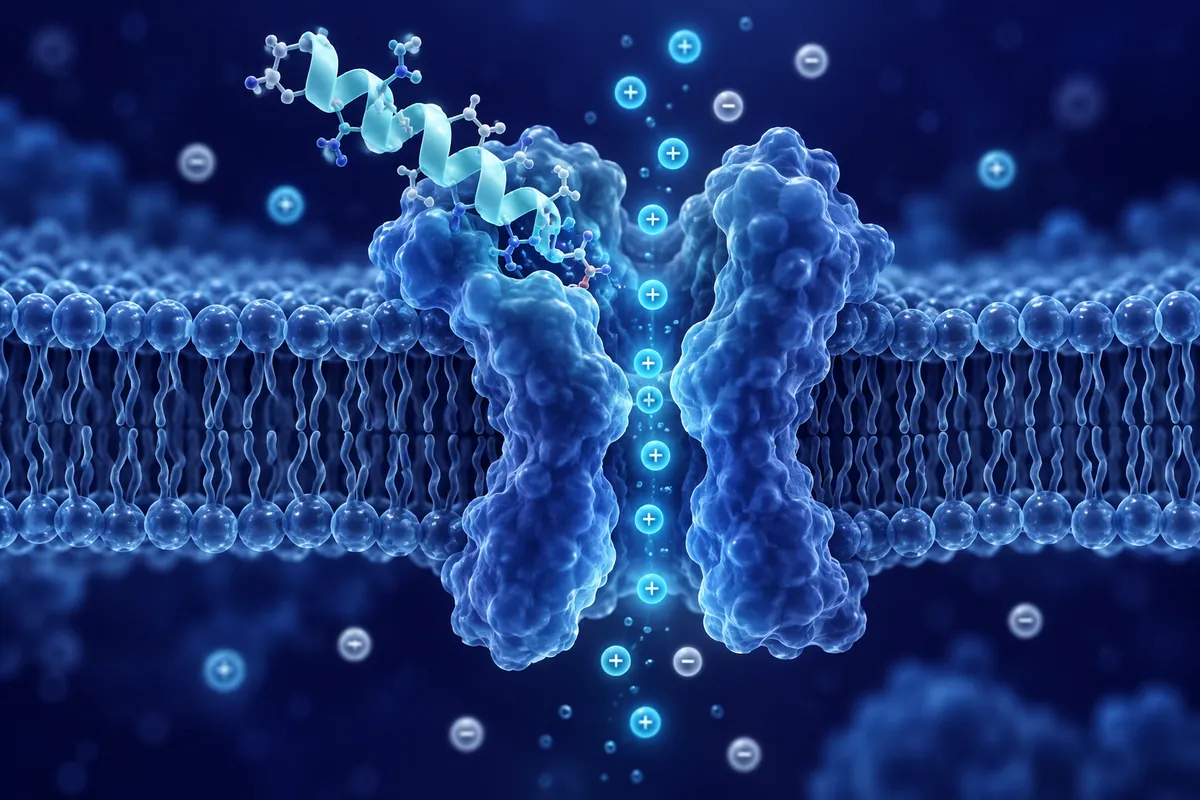
PNA’s charge neutrality, while beneficial for binding stability, creates a delivery problem. Without negative charge, PNAs do not interact productively with cationic lipid transfection reagents designed for nucleic acid delivery. Cellular uptake of unmodified PNAs is poor : they do not cross cell membranes efficiently on their own.
Researchers have addressed this through conjugation strategies. Cell-penetrating peptides (CPPs) : another peptide research category : are frequently attached to PNA sequences to facilitate cellular entry. The resulting CPP-PNA conjugates combine the membrane-penetrating ability of the CPP with the sequence-specific binding of the PNA. Other delivery approaches include encapsulation in nanoparticles, conjugation to receptor-targeting ligands, and electroporation for in vitro applications.
Research Applications Beyond Gene Silencing
PNA technology has expanded beyond its original antisense/antigene applications. FISH (fluorescence in situ hybridization) probes built from PNA sequences offer faster hybridization kinetics and higher signal-to-noise ratios than DNA-based FISH probes, particularly for repetitive sequences. PNA molecular beacons : hairpin-structured probes that fluoresce upon target binding : show improved sensitivity for real-time detection assays.
In biosensor research, PNA capture probes immobilized on electrode surfaces detect complementary nucleic acid sequences with picomolar sensitivity. The neutral backbone reduces non-specific electrostatic interactions with the sensor surface, improving signal specificity compared to charged DNA probes.
More recently, PNA-based genome editing tools have emerged as alternatives to CRISPR in certain research contexts. Triplex-forming PNAs can stimulate site-specific recombination when delivered with donor DNA templates, producing targeted gene modifications without nuclease-induced double-strand breaks : and therefore without the off-target cleavage risks associated with nuclease-based approaches.
Disclaimer: This content is intended for research purposes only and is not meant to constitute medical advice.
This educational material discusses laboratory research concepts only. Loti Labs products are intended strictly for research and development use and are not for human consumption.