A Hormone from Bone
For most of its research history, osteocalcin was considered a simple structural protein : one of many matrix molecules embedded in mineralized bone tissue during formation. Produced exclusively by osteoblasts and encoded by the BGLAP gene, this 49-amino-acid peptide was studied primarily as a serum biomarker of bone turnover. Then Gerard Karsenty’s laboratory at Columbia University upended that narrative entirely.
In a landmark 2007 publication, Karsenty’s group demonstrated that osteocalcin knockout mice were not just osteoporotic : they were also obese, glucose-intolerant, and had reduced pancreatic beta-cell mass. The skeleton was not merely a passive framework. It was an endocrine organ, and osteocalcin was its hormone.
Two Forms, Two Functions
Osteocalcin exists in two forms distinguished by a post-translational modification. Carboxylated osteocalcin (cOCN) carries gamma-carboxyglutamic acid (Gla) residues at positions 17, 21, and 24, created by a vitamin K-dependent carboxylase. These Gla residues bind calcium ions with high affinity, anchoring carboxylated osteocalcin to the hydroxyapatite mineral phase of bone matrix. This is the structural form : embedded in bone and serving a matrix role.
Undercarboxylated osteocalcin (ucOCN) lacks some or all Gla residues and has correspondingly reduced affinity for hydroxyapatite. During bone resorption, osteoclasts acidify the resorption lacuna, and the low pH decarboxylates matrix-bound osteocalcin, releasing the undercarboxylated form into the bloodstream. This ucOCN is the endocrine form : the fraction that travels through circulation and acts on distant organs.
This resorption-dependent activation mechanism is elegant. It couples bone turnover directly to metabolic signaling: when bone is being actively remodeled (high osteoclast activity), more ucOCN enters circulation, amplifying metabolic effects. When remodeling is quiescent, less ucOCN is released. The skeleton effectively broadcasts its remodeling status to the rest of the body.
Glucose Metabolism and Pancreatic Function
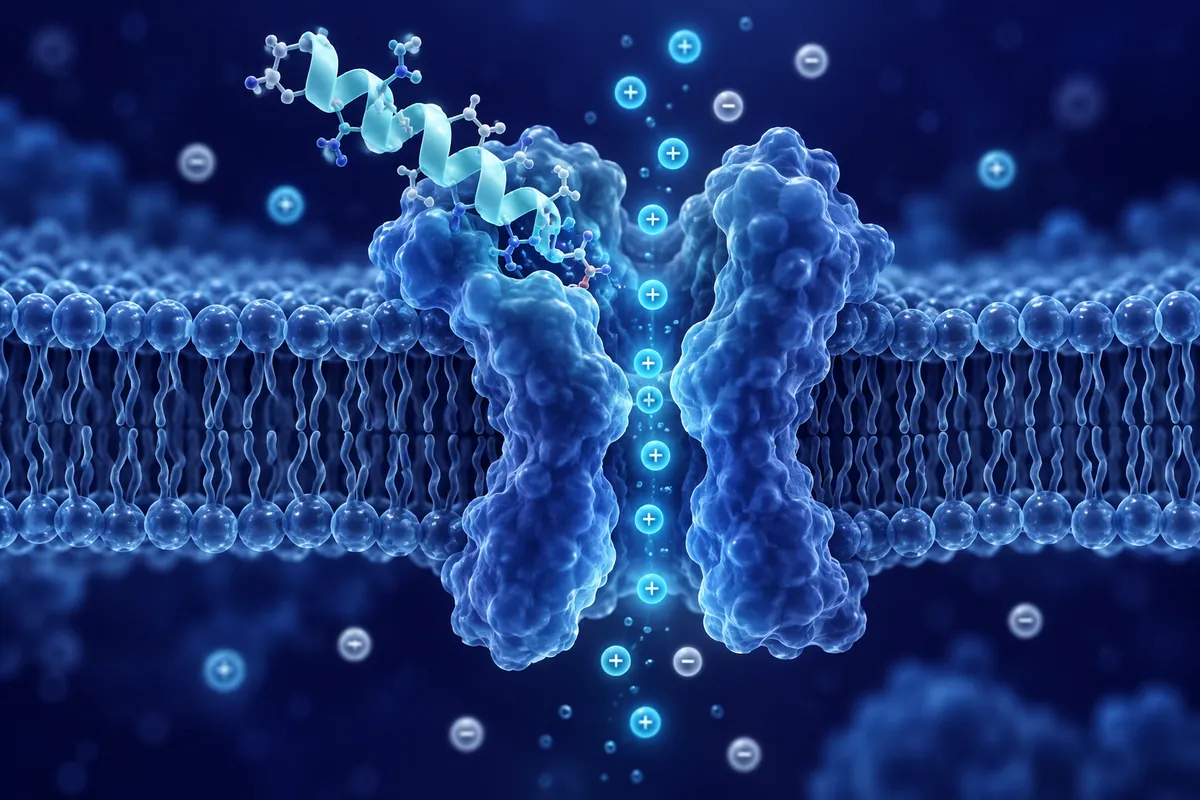
The metabolic axis of osteocalcin signaling operates through GPRC6A, a G-protein-coupled receptor expressed on pancreatic beta-cells, adipocytes, hepatocytes, and Leydig cells. In pancreatic beta-cells, ucOCN binding to GPRC6A stimulates insulin expression and secretion through a cAMP-dependent pathway.
The evidence from mouse genetics is compelling. Osteocalcin-deficient mice show reduced beta-cell proliferation, decreased insulin secretion in response to glucose challenge, and impaired glucose tolerance. Conversely, infusion of recombinant ucOCN in wild-type mice improves glucose disposal and increases insulin sensitivity in adipose tissue and muscle : effects mediated at least partly through increased adiponectin production from fat cells.
The osteocalcin-adiponectin connection creates a feedforward loop between bone and fat. ucOCN stimulates adiponectin release from adipocytes. Adiponectin, in turn, enhances insulin sensitivity in liver and muscle. Meanwhile, insulin signaling in osteoblasts promotes osteocalcin production and osteoclast-mediated decarboxylation. Bone, fat, and pancreas communicate in a triangular endocrine circuit that none of these organs was previously thought to participate in.
Cognitive Function: The Bone-Brain Axis
Perhaps the most surprising chapter in the osteocalcin story involves the brain. In 2013, Karsenty’s group reported that osteocalcin crosses the blood-brain barrier and influences neurotransmitter synthesis in the hippocampus and brainstem. Osteocalcin-null mice showed increased anxiety-like behavior and impaired spatial learning in Morris water maze tests.
The mechanism involves direct promotion of monoamine neurotransmitter synthesis. ucOCN binding to GPRC6A (or a related receptor) in brainstem raphe nuclei and locus coeruleus neurons upregulates tryptophan hydroxylase 2 (serotonin synthesis) and tyrosine hydroxylase (dopamine and norepinephrine synthesis). More neurotransmitter precursor enzymes means more serotonin, dopamine, and norepinephrine available for synaptic signaling.
In hippocampal neurons, osteocalcin enhances BDNF expression and promotes dendritic arborization in culture. These effects parallel those reported for exercise-induced irisin signaling, raising the intriguing possibility that multiple exercise-related hormones : from both muscle and bone : converge on hippocampal plasticity through complementary mechanisms.
Exercise and Osteocalcin Release
Physical exercise increases circulating ucOCN through two mechanisms. Mechanical loading directly stimulates osteoblast osteocalcin production. Simultaneously, exercise-induced acidosis and increased bone remodeling promote osteoclastic decarboxylation of stored matrix osteocalcin. Together, these produce acute exercise-related spikes in circulating ucOCN.
A 2019 study by Mera and colleagues demonstrated that osteocalcin mediates some of the acute exercise capacity effects in aging mice. The mechanism involved osteocalcin-stimulated glucose and fatty acid uptake in muscle fibers during sustained effort : a metabolic support function previously unattributed to any bone-derived factor.
Male Fertility Connection
Adding another dimension, ucOCN acts on Leydig cells in the testes through GPRC6A, stimulating testosterone biosynthesis. Osteocalcin-null male mice show reduced testis size and testosterone concentrations, along with decreased fertility. This bone-gonad axis was completely unexpected and suggested that the skeleton participates in reproductive endocrinology : a paradigm shift for a tissue long considered hormonally inert.
Aging and Decline
Circulating ucOCN concentrations decline with aging in rodent models, a decline that parallels age-related changes researchers have mapped to glucose tolerance, cognitive, muscle, and gonadal endpoints in animal work : the range of tissues where osteocalcin signaling has been shown to act. Whether declining osteocalcin is a driver or merely a correlate of these age-related changes remains an active area of investigation, and restoring youthful osteocalcin signaling in aging rodent models continues to be explored as a research strategy.
Disclaimer: This content is intended for research purposes only and is not meant to constitute medical advice.
This educational material discusses laboratory research concepts only. Loti Labs products are intended strictly for research and development use and are not for human consumption.