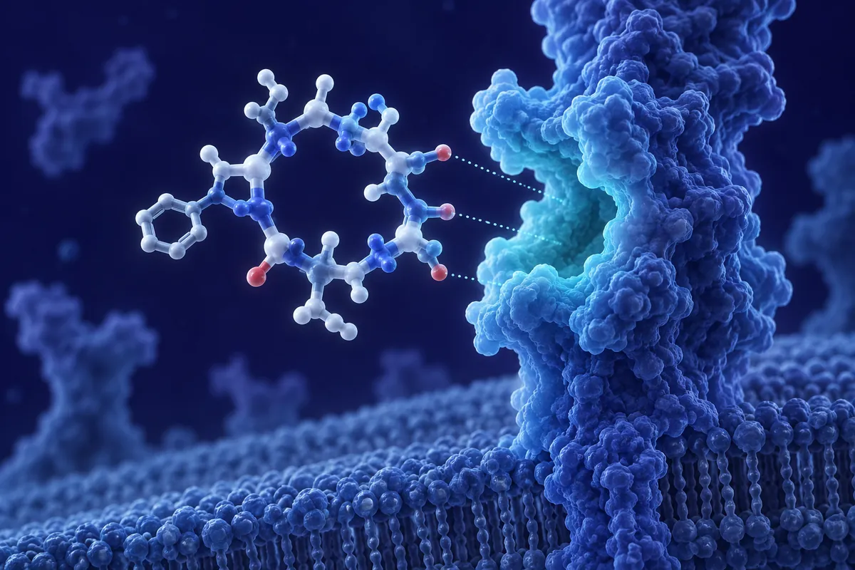
Alpha-MSH has always been a strange molecule to study. Cut it down to just three residues : Lysine-Proline-Valine, or KPV : and something curious happens: the pigmentation signaling drops away, but the anti-inflammatory activity sticks around almost fully intact. That dissociation is the whole reason KPV has drawn sustained interest from researchers working in mucosal immunology, dermatologic inflammation models, and gut barrier research over the past two decades.
Why would a fragment this small retain any biological activity at all? Melanocortin peptides are built from a core pharmacophore, and KPV happens to sit right at the C-terminal tripeptide of alpha-MSH, a sequence long recognized as a minimal active site for certain non-pigmentary effects. Researchers investigating this fragment have found it behaves less like a classical melanocortin receptor agonist and more like a signaling modulator working through alternative, still-debated pathways.
The Melanocortin Receptor Question
Full-length alpha-MSH acts primarily through melanocortin receptors 1 and 4 (MC1R, MC4R), the former driving melanogenesis in melanocytes and the latter tied to energy homeostasis in hypothalamic circuits. KPV, notably, shows weak to negligible binding affinity at these receptors in several in vitro binding assays. That is precisely the point researchers keep returning to.
Because KPV exerts anti-inflammatory effects independent of classical MC1R/MC4R engagement, investigators have proposed it may modulate NF-κB signaling directly at the intracellular level, bypassing canonical receptor-ligand docking. Some published cell-culture work has reported that KPV can suppress IL-1β and TNF-α-induced NF-κB nuclear translocation in intestinal epithelial cell lines, an effect measured independently of any detectable melanocortin receptor expression on those cells.
Mucosal and Gut Barrier Models
A meaningful share of KPV research has centered on colonic epithelium. In rodent models of induced colitis : often using dextran sulfate sodium (DSS) or trinitrobenzene sulfonic acid (TNBS) protocols : KPV administration has been associated with reduced histological damage scores and lower pro-inflammatory cytokine expression in colonic tissue homogenates. Several of these studies also measured effects on tight junction protein expression, including zonula occludens-1 (ZO-1) and occludin, both central to intestinal barrier integrity.
What makes this line of research particularly interesting is the dose-response pattern researchers have reported: efficacy in these colitis models often plateaus at relatively low concentrations, consistent with a receptor-independent or highly efficient signaling mechanism rather than a simple mass-action effect.
Keratinocyte and Skin Inflammation Work
Parallel research has explored KPV in keratinocyte culture systems and topical inflammation models. Ultraviolet-induced inflammatory markers, along with cytokine cascades relevant to atopic-type skin inflammation, have been reported to attenuate with KPV exposure in several in vitro systems. Because keratinocytes do express some melanocortin receptor subtypes, this line of work has actually helped researchers tease apart which KPV effects are receptor-mediated versus which persist even when receptor expression is minimal or pharmacologically blocked.
Stability and the Structure-Activity Puzzle
Small peptides face an obvious challenge: proteolytic degradation. KPV’s three-residue length makes it vulnerable to rapid breakdown by peptidases in physiological environments, and early pharmacokinetic work noted a fairly short half-life in plasma-like conditions. This has pushed structure-activity relationship (SAR) researchers toward D-amino acid substitutions and cyclization strategies, aiming to preserve the anti-inflammatory pharmacophore while extending stability in research models.
Is a tripeptide really the minimal unit, or could even smaller modifications retain function? That question keeps SAR labs occupied. Some analog work has substituted the central proline with structurally related residues to probe conformational requirements, with mixed results on retained activity : a reminder that even minimal peptides encode real structural specificity, not just amino acid composition.
Open Mechanistic Questions
- Whether KPV acts through an as-yet uncharacterized receptor rather than pure receptor-independent signaling
- How KPV’s effects on macrophage polarization (M1 to M2 shift) relate to its epithelial actions
- Whether systemic versus local delivery routes meaningfully change the inflammatory signaling profile observed
Researchers have also looked at KPV’s interaction with toll-like receptor signaling cascades, given the overlap between TLR4 activation and the NF-κB pathways KPV appears to modulate. This intersection is still being mapped out, and conflicting findings across different cell lines suggest the mechanism may not be uniform across tissue types.
Where the Research Goes Next
KPV occupies an unusual niche: small enough to synthesize cheaply and modify extensively, yet biologically active enough to keep mucosal immunology and dermatologic inflammation labs interested. Future research will likely lean harder into receptor identification studies, using knockout models and radioligand competition assays to settle whether an unrecognized binding partner exists. Combination work pairing KPV with other melanocortin fragments, or testing it alongside established anti-inflammatory research agents, may also clarify whether its effects are additive or mechanistically distinct.
For now, the tripeptide remains a compact case study in how much biological information can be packed into three amino acids : and how much is still left to figure out about where that information goes once it enters a cell.
There is also growing interest in how KPV research might intersect with broader melanocortin system mapping. MC3R, a receptor subtype expressed heavily in macrophages and other immune cells, has emerged as another candidate worth testing against KPV binding, given that MC1R and MC4R have largely been ruled out as primary mediators. If MC3R plays even a partial role, that would reshape how researchers interpret decades of prior KPV data collected without that subtype in mind.
Comparative work against other short melanocortin fragments has also appeared in the literature. Some groups have tested KPV alongside alpha-MSH(11-13) analogs bearing subtle side-chain modifications, looking for a fragment that retains anti-inflammatory potency while gaining proteolytic resistance beyond what D-amino acid substitution alone provides. Early results are mixed, but the comparative approach is useful: it helps separate which structural features are essential from which are merely conserved by accident.
None of this diminishes how unusual KPV already is as a research subject. A three-residue peptide that modulates inflammatory signaling without engaging the receptors its parent molecule depends on is not a common finding, and it keeps inviting follow-up work because the mechanism isn’t fully settled.
Disclaimer: This content is intended for research purposes only and is not meant to constitute medical advice.
This educational material discusses laboratory research concepts only. Loti Labs products are intended strictly for research and development use and are not for human consumption.