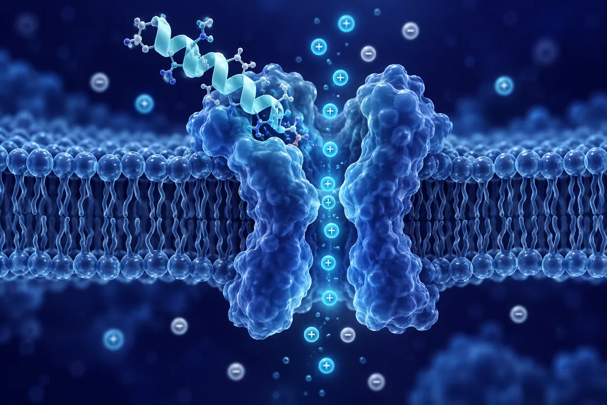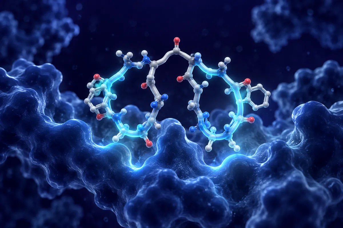
Alpha-melanocyte-stimulating hormone (α-MSH) is a 13-amino-acid peptide derived from the post-translational processing of proopiomelanocortin (POMC) in the anterior pituitary, hypothalamus, and various peripheral tissues. But that is barely the opening act.
The melanocortin system comprises five receptor subtypes (MC1R through MC5R), and α-MSH activates most of them.
Binding to MC1R on macrophages triggers a cAMP-dependent cascade that suppresses NF-κB nuclear translocation. The effect is robust and reproducible across multiple cell lines and primary cultures.
But MC1R is not alone. The effect was abolished in MC3R knockout animals, confirming receptor specificity.
What distinguishes α-MSH from conventional immunosuppressants in preclinical research is selectivity.
POMC neurons in the arcuate nucleus release α-MSH in response to leptin signaling, which then activates MC4R on second-order neurons in the paraventricular nucleus.
This pathway’s importance was demonstrated dramatically by genetic studies. MC4R knockout mice develop severe obesity. In humans, MC4R is the most commonly mutated gene in monogenic obesity : heterozygous loss-of-function mutations affect approximately 5-6% of individuals with severe early-onset obesity.
Intriguingly, agouti-related peptide (AgRP) acts as an endogenous antagonist at MC4R, creating a push-pull system. The balance between α-MSH and AgRP at the MC4R determines the net orexigenic or anorexigenic signal. Researchers manipulating this balance in rodent models have achieved bidirectional control of feeding behavior with remarkable precision.
Aqueous humor contains measurable concentrations of α-MSH, where it suppresses antigen-presenting cell activation and T-cell proliferation.
This ocular research has broader implications.
Melanocortin receptors are expressed in cardiac tissue and vascular endothelium. In isolated heart preparations and perfused vascular beds, α-MSH produces vasodilation through nitric oxide (NO)-dependent mechanisms.
Structural Features and Stability
α-MSH (Ac-Ser-Tyr-Ser-Met-Glu-His-Phe-Arg-Trp-Gly-Lys-Pro-Val-NH₂) carries an N-terminal acetyl group and C-terminal amidation : modifications that enhance its resistance to aminopeptidase and carboxypeptidase degradation compared to unmodified POMC fragments. Even so, its plasma half-life is short, measured in minutes rather than hours.
This instability drove the development of synthetic melanocortin analogs like NDP-α-MSH (Melanotan I), which substitutes norleucine for methionine at position 4 and D-phenylalanine for L-phenylalanine at position 7. These modifications dramatically extend half-life and increase receptor binding affinity, making NDP-α-MSH a valuable pharmacological tool for research : though it is a distinct compound from native α-MSH with different receptor selectivity profiles.
Open Research Questions
Several frontiers remain. How does α-MSH signaling change across the lifespan? POMC neuron activity appears to decline with aging in rodent models, potentially contributing to age-related metabolic shifts. Does peripheral α-MSH production in keratinocytes, gut epithelium, and immune cells serve purely local (autocrine/paracrine) functions, or does it contribute meaningfully to systemic melanocortin tone? Receptor subtype-selective analogs may hold the answer, but selectivity at the required level remains a design challenge in peptide chemistry.
Disclaimer: This content is intended for research purposes only and is not meant to constitute medical advice.
This educational material discusses laboratory research concepts only. Loti Labs products are intended strictly for research and development use and are not for human consumption.