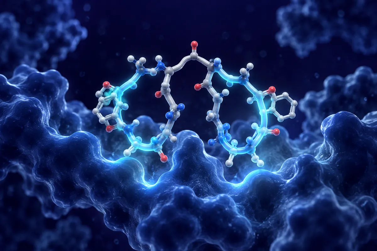
Catestatin isn’t a new name. It has been sitting in the neuroendocrine literature since the late 1990s, mostly filed under blood pressure and catecholamine release. So why are neuroscience labs paying attention to it now?
The short answer is tau. A September 2026 Cell Press report looked at the catestatin peptide in mouse models of tauopathy and amyloid pathology, and it linked adrenergic signaling to the way these proteins build up in the brain. That’s a different frame from the one most researchers associate with this fragment, and it deserves a careful look.
Where Catestatin Comes From
Chromogranin A (CgA) is an acidic protein packed into the secretory granules of neurons and endocrine cells. It’s abundant. It also gets cut up, and the pieces have their own activities.
Proteases such as prohormone convertases and cathepsins process CgA into several fragments, including vasostatin, pancreastatin and catestatin. Catestatin corresponds to a 21-residue stretch near the C-terminal region of the parent protein (often written as CgA352-372 for the human sequence). Bovine, rat and mouse versions differ at a few positions, which matters if you are matching a peptide to a rodent model.
Its best known property, worked out in cell systems like PC12 and bovine chromaffin cells, is that it blocks nicotinic acetylcholine receptors. That block dampens catecholamine secretion. In other words, it acts as a kind of brake on the adrenergic system that released it in the first place. A neat feedback loop, if you like that sort of thing.
Variants worth knowing
Naturally occurring sequence variants (for example Gly364Ser and Pro370Leu in the human sequence) have been characterized in vitro, and they differ in how strongly they inhibit nicotinic signaling. For researchers, that variation is useful. It gives you built-in comparison peptides with slightly different potencies.
Why Adrenergic Signaling Matters for Tau and Amyloid
The locus coeruleus, a small brainstem nucleus, supplies most of the forebrain’s noradrenaline. It’s also one of the earliest regions where abnormal tau shows up in post-mortem staging work. That anatomical overlap has kept noradrenergic signaling on the radar for a long time.
In rodent and cell studies, adrenergic receptors, especially beta-2 and alpha-2A subtypes, have been tied to amyloid precursor protein processing and to kinase pathways that phosphorylate tau. The picture is messy. Some studies find that blocking a receptor reduces pathology, while others find the opposite depending on the model, the age of the animals and the receptor subtype. Anyone who has tried to reconcile this literature knows the feeling.
Catestatin offers a different angle. Rather than targeting one receptor subtype with a small molecule, it acts upstream by limiting catecholamine output, and it seems to interact with immune signaling as well. That’s what makes the September work interesting to us.
What the Mouse Model Work Reported
The study used transgenic mouse lines that develop tau pathology and amyloid deposits. The team examined CgA and catestatin levels in these models and then tested what happened when catestatin signaling was changed.
Their reported findings point toward reduced adrenergic tone being associated with less tau phosphorylation and lower amyloid burden in the treated rodent groups compared with controls. Microglial activation markers also shifted. We’d encourage readers to go to the paper itself for the exact figures, since effect sizes vary across brain regions and readouts, and summarizing them loosely would only blur the picture.
A few design questions stand out. How was the peptide delivered to the brain, and how much crossed the blood-brain barrier? Were sex differences examined? Did the changes persist after administration stopped? These are the details that decide whether a result is reproducible or just a single-lab curiosity.
Interpreting it cautiously
Mouse tauopathy lines overexpress mutant human tau, which accelerates pathology in ways that don’t always mirror sporadic disease biology. Amyloid models have similar limits. Results from them are a starting point. They’re hypothesis generating, not settled facts.
Practical Considerations for Lab Work
Catestatin is a fairly cationic peptide, rich in arginine. That charge helps with membrane interactions and probably explains part of its antimicrobial activity seen in vitro, but it can also cause sticking to plastics and glass. Low-binding tubes help. So does checking concentration after reconstitution instead of assuming it.
Species matching deserves attention too. If your model is a mouse, the mouse sequence is the natural choice, although many published studies have used the human or bovine version. Note which one you used. Reviewers will ask.
Controls are where a lot of peptide studies fall short. A scrambled sequence with the same composition, a vehicle group and, ideally, a variant peptide with known lower potency make the results much easier to interpret. Measuring catecholamine levels directly, rather than inferring them, closes another gap.
Then there’s timing. Tau pathology develops over months in most lines, so the age at which administration begins can change the outcome completely. Early intervention studies and late ones answer different questions.
Open Questions
Several things remain unclear. Does catestatin act mainly through nicotinic receptor blockade in the brain, or are its immune effects on microglia doing more of the work? Is the relevant source peripheral, central, or both? And how do the other CgA fragments, some of which push in opposite directions, fit into the same story?
There’s also the question of the parent protein. CgA itself is elevated in some neurodegeneration models, and its processing may change with age. Measuring the ratio of intact CgA to catestatin could turn out to be more informative than tracking either one alone.
Conclusion
Catestatin research has moved well beyond its cardiovascular roots. The September 2026 mouse data connect a modest 21-residue fragment to tau and amyloid pathology through adrenergic and immune pathways, which is a genuinely interesting link. It is still early. Replication in other model systems, careful brain delivery measurements and clean controls will decide how far this line of inquiry goes. For labs already working on locus coeruleus biology, though, it’s a peptide worth adding to the toolkit.
Disclaimer: This content is intended for research purposes only and is not meant to constitute medical advice.
This educational material discusses laboratory research concepts only. Loti Labs products are intended strictly for research and development use and are not for human consumption.