What Is Irisin?
Irisin burst onto the research scene in 2012 when a team led by Bruce Spiegelman at Harvard identified it as a cleaved fragment of fibronectin type III domain-containing protein 5 (FNDC5). That single observation opened an entirely new chapter in exercise biology.
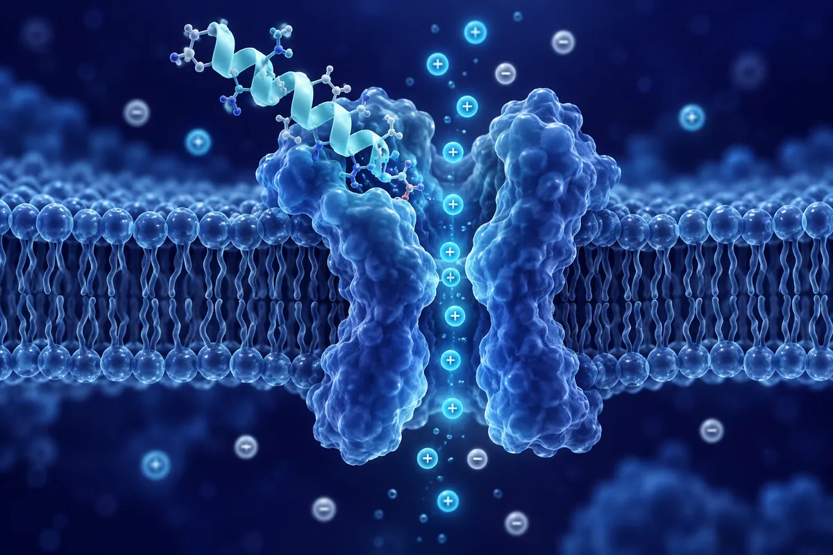
At just 112 amino acids, irisin is classified as a myokine : a signaling molecule secreted by muscle fibers. Its name comes from Iris, the Greek messenger goddess, reflecting the peptide’s role as a long-range endocrine courier between muscle and distant tissues.
The Browning Mechanism: White Fat to Beige Fat
White adipocytes store energy. Brown adipocytes burn it.
How does it work? Irisin activates p38 MAPK and ERK signaling cascades in precursor adipocytes, upregulating peroxisome proliferator-activated receptor gamma coactivator 1-alpha (PGC-1α) and uncoupling protein 1 (UCP1).
Muscle and bone are mechanical neighbors, but irisin revealed a biochemical conversation between them that few anticipated. In osteoblast cultures, recombinant irisin stimulates alkaline phosphatase activity and mineralization through activation of the integrin αV/β5 receptor complex.
Simultaneously, irisin appears to suppress osteoclast differentiation.
Perhaps the most intriguing irisin research has emerged from neuroscience laboratories. FNDC5/irisin expression has been confirmed in multiple brain regions, including the hippocampus, Purkinje cells, and hypothalamus.
The mechanism appears to involve brain-derived neurotrophic factor (BDNF).
Receptor Biology and Signaling Partners
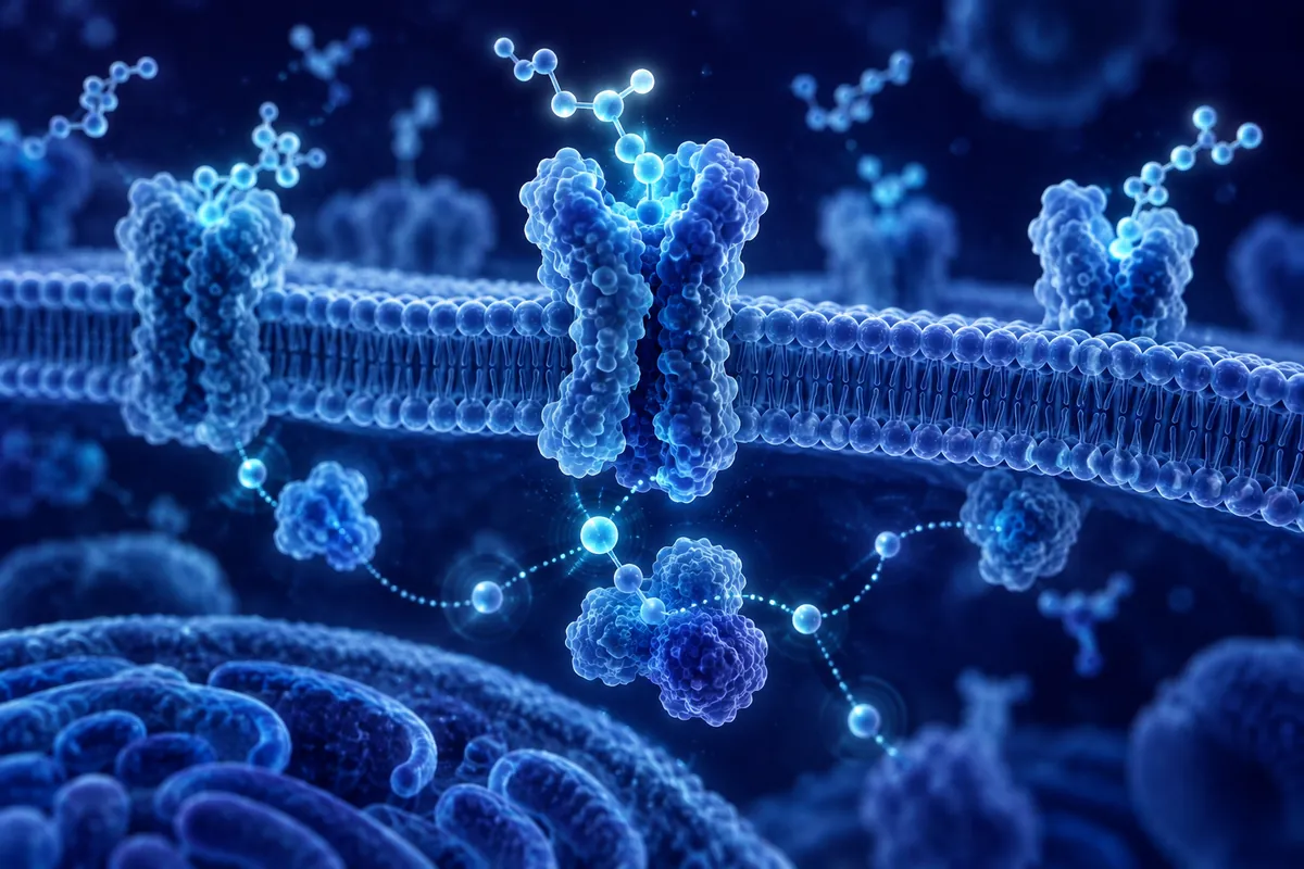
Identifying irisin’s receptor proved surprisingly difficult. In 2018, researchers confirmed that irisin binds to integrin αVβ5 on osteocytes with nanomolar affinity. Subsequent work extended this to integrin αVβ1 in adipose tissue.
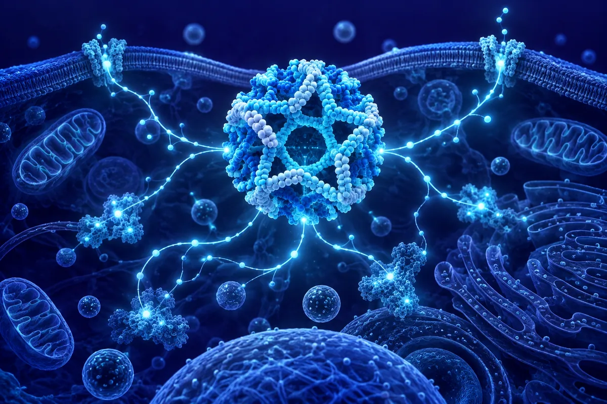
Downstream signaling varies by tissue type. In adipocytes, irisin primarily activates AMPK. In neurons, the ERK-CREB cascade dominates. In bone, β-catenin takes the lead. This context-dependent signaling profile makes irisin a particularly rich subject for systems biology approaches, where researchers map how a single peptide ligand produces tissue-specific outcomes through shared but differently wired intracellular networks.
Exercise Intensity and Irisin Release
Not all exercise is equal when it comes to irisin secretion. Studies measuring circulating irisin concentrations in response to different exercise protocols have consistently shown that high-intensity resistance training produces the largest acute spikes, followed by high-intensity interval training (HIIT), with moderate steady-state cardio producing more modest elevations.
The kinetics are rapid. Circulating irisin peaks approximately 30-60 minutes post-exercise and returns to baseline within two to three hours. This pulsatile release pattern mirrors other exercise-induced myokines and distinguishes irisin from constitutively secreted hormones. Researchers studying irisin’s downstream effects must account for this transient exposure profile when designing in vivo protocols.
Current Research Frontiers
Several open questions drive current irisin research forward. First, the quantification debate: some early ELISA-based assays for circulating irisin lacked specificity, producing inflated baseline values. Mass spectrometry-based methods developed after 2015 provided more reliable measurements and confirmed that circulating irisin does increase with exercise, though at lower absolute concentrations than initially reported.
Second, the sarcopenia angle. As skeletal muscle mass declines with aging, irisin secretion capacity may fall in parallel. Whether this decline contributes to age-related metabolic shifts, bone density changes, and cognitive decline : or merely correlates with them : is a question that longitudinal research is beginning to address.
Third, the interplay between irisin and other myokines. Muscle releases hundreds of signaling molecules during contraction. Understanding how irisin interacts with IL-6, myostatin, meteorin-like, and other exercise factors may reveal synergistic or antagonistic relationships with implications for exercise physiology research.
Disclaimer: This content is intended for research purposes only and is not meant to constitute medical advice.
This educational material discusses laboratory research concepts only. Loti Labs products are intended strictly for research and development use and are not for human consumption.